1ZT4 | pdb_00001zt4

The crystal structure of human CD1d with and without alpha-Galactosylceramide


Experimental Data Snapshot

  • Method: X-RAY DIFFRACTION
  • Resolution: 3.00 Å
  • R-Value Free: 
    0.326 (Depositor), 0.317 (DCC) 
  • R-Value Work: 
    0.234 (Depositor), 0.229 (DCC) 
  • R-Value Observed: 
    0.234 (Depositor) 

Starting Model: experimental
View more details

wwPDB Validation 3D Report Full Report

Validation slider image for 1ZT4

Ligand Structure Quality Assessment 


This is version 2.2 of the entry. See complete history

Literature

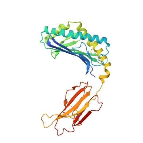
The crystal structure of human CD1d with and without alpha-galactosylceramide

Koch, M.Stronge, V.S.Shepherd, D.Gadola, S.D.Mathew, B.Ritter, G.Fersht, A.R.Besra, G.S.Schmidt, R.R.Jones, E.Y.Cerundolo, V.

(2005) Nat Immunol 6: 819-826

  • DOI: https://doi.org/10.1038/ni1225
  • Primary Citation Related Structures: 
    1ZT4

  • PubMed Abstract: 

    The glycolipid alpha-galactosylceramide binds with high affinity to CD1d and stimulates natural killer T cells. Here we report the crystal structure of human CD1d in complex with synthetic alpha-galactosylceramide at a resolution of 3.0 A. The structure shows a tightly fit lipid in the CD1d binding groove, with the sphingosine chain bound in the C' pocket and the longer acyl chain anchored in the A' pocket. We also present the CD1d structure without lipid, which has a more open conformation of the binding groove, suggesting a dual conformation of CD1d in which the 'open' conformation is more able to load lipids. These structures provide clues as to how CD1 molecules load glycolipids as well as data to guide the design of new therapeutic agents.


  • Organizational Affiliation
    • Cancer Research UK Receptor Structure Research Group, The Henry Wellcome Building for Genomic Medicine, Roosevelt Drive, Headington, Oxford OX3 7BN, UK.

Macromolecule Content 

  • Total Structure Weight: 88.25 kDa 
  • Atom Count: 6,093 
  • Modeled Residue Count: 750 
  • Deposited Residue Count: 762 
  • Unique protein chains: 2

Macromolecules

Find similar proteins by:|  3D Structure
Entity ID: 1
MoleculeChains  Sequence LengthOrganismDetailsImage
T-cell surface glycoprotein CD1d
A, C
281Homo sapiensMutation(s): 0 
Gene Names: CD1D
UniProt & NIH Common Fund Data Resources
Find proteins for P15813 (Homo sapiens)
Explore P15813 
Go to UniProtKB:  P15813
PHAROS:  P15813
GTEx:  ENSG00000158473 
Entity Groups
Sequence Clusters30% Identity50% Identity70% Identity90% Identity95% Identity100% Identity
UniProt GroupP15813
Sequence Annotations
Expand
Reference Sequence
Find similar proteins by:|  3D Structure
Entity ID: 2
MoleculeChains  Sequence LengthOrganismDetailsImage
Beta-2-microglobulin
B, D
100Homo sapiensMutation(s): 0 
Gene Names: B2M
UniProt & NIH Common Fund Data Resources
Find proteins for P61769 (Homo sapiens)
Explore P61769 
Go to UniProtKB:  P61769
PHAROS:  P61769
GTEx:  ENSG00000166710 
Entity Groups
Sequence Clusters30% Identity50% Identity70% Identity90% Identity95% Identity100% Identity
UniProt GroupP61769
Sequence Annotations
Expand
Reference Sequence

Small Molecules

Ligands 1 Unique
IDChains Name / Formula / InChI Key2D Diagram3D Interactions
AGH

Query on AGH


Download:Ideal Coordinates CCD File
E [auth A]N-{(1S,2R,3S)-1-[(ALPHA-D-GALACTOPYRANOSYLOXY)METHYL]-2,3-DIHYDROXYHEPTADECYL}HEXACOSANAMIDE
C50 H99 N O9
VQFKFAKEUMHBLV-BYSUZVQFSA-N

Experimental Data & Validation

Experimental Data

  • Method: X-RAY DIFFRACTION
  • Resolution: 3.00 Å
  • R-Value Free:  0.326 (Depositor), 0.317 (DCC) 
  • R-Value Work:  0.234 (Depositor), 0.229 (DCC) 
  • R-Value Observed: 0.234 (Depositor) 
Space Group: P 21 21 21
Unit Cell:
Length ( Å )Angle ( ˚ )
a = 50.47α = 90
b = 94.3β = 90
c = 176.07γ = 90
Software Package:
Software NamePurpose
CNSrefinement
PDB_EXTRACTdata extraction
DENZOdata reduction
SCALEPACKdata scaling
AMoREphasing
REFMACrefinement

Structure Validation

View Full Validation Report


Ligand Structure Quality Assessment 


Entry History 

Revision History  (Full details and data files)

  • Version 1.0: 2005-07-19
    Type: Initial release
  • Version 1.1: 2007-10-16
    Changes: Version format compliance
  • Version 1.2: 2011-07-13
    Changes: Derived calculations, Version format compliance
  • Version 1.3: 2019-07-24
    Changes: Data collection, Refinement description
  • Version 2.0: 2020-06-17
    Changes: Data collection, Database references, Polymer sequence
  • Version 2.1: 2023-08-23
    Changes: Data collection, Database references, Derived calculations, Refinement description
  • Version 2.2: 2024-11-20
    Changes: Structure summary