1ZLQ | pdb_00001zlq

Crystallographic and spectroscopic evidence for high affinity binding of Fe EDTA (H2O)- to the periplasmic nickel transporter NikA


Experimental Data Snapshot

  • Method: X-RAY DIFFRACTION
  • Resolution: 1.80 Å
  • R-Value Free: 
    0.218 (Depositor) 
  • R-Value Work: 
    0.163 (Depositor), 0.165 (DCC) 
  • R-Value Observed: 
    0.165 (Depositor) 

Starting Model: experimental
View more details

wwPDB Validation 3D Report Full Report

Validation slider image for 1ZLQ

Ligand Structure Quality Assessment 


This is version 1.4 of the entry. See complete history

Literature

Crystallographic and Spectroscopic Evidence for High Affinity Binding of FeEDTA(H(2)O)(-) to the Periplasmic Nickel Transporter NikA

Cherrier, M.V.Martin, L.Cavazza, C.Jacquamet, L.Lemaire, D.Gaillard, J.Fontecilla Camps, J.C.

(2005) J Am Chem Soc 127: 10075-10082

  • DOI: https://doi.org/10.1021/ja0518530
  • Primary Citation Related Structures: 
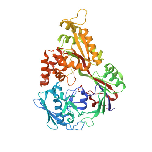
    1ZLQ

  • PubMed Abstract: 

    Because nickel is both essential and toxic to a great variety of organisms, its detection and transport is highly regulated. In Escherichia coli and other related Gram-negative bacteria, high affinity nickel transport depends on proteins expressed by the nik operon. A central actor of this process is the periplasmic NikA transport protein. A previous structural report has proposed that nickel binds to NikA as a pentahydrate species. However, both stereochemical considerations and X-ray absorption spectroscopic results are incompatible with that interpretation. Here, we report the 1.8 A resolution structure of NikA and show that it binds FeEDTA(H2O)- with very high affinity. In addition, we provide crystallographic evidence that a metal-EDTA complex was also bound to the previously reported NikA structure. Our observations strongly suggest that nickel transport in E. coli requires the binding of this metal ion to a metallophore that bears significant resemblance to EDTA. They also provide a basis for the potential use of NikA in the bioremediation of toxic transition metals and the design of artificial metalloenzymes.


  • Organizational Affiliation
    • Laboratoire de Cristallographie et de Cristallogenèse des Protéines and Laboratoire de Spectroscopie de Masse des Protéines, Institut de Biologie Structurale J.P. Ebel (CEA-CNRS-UJF), 41 rue Jules Horowitz, 38027 Grenoble Cedex 1, France.

Macromolecule Content 

  • Total Structure Weight: 114.84 kDa 
  • Atom Count: 9,116 
  • Modeled Residue Count: 998 
  • Deposited Residue Count: 1,004 
  • Unique protein chains: 1

Macromolecules

Find similar proteins by:|  3D Structure
Entity ID: 1
MoleculeChains  Sequence LengthOrganismDetailsImage
Nickel-binding periplasmic protein
A, B
502Escherichia coliMutation(s): 0 
Gene Names: nikA
EC: 3.6.3.24
UniProt
Find proteins for P33590 (Escherichia coli (strain K12))
Explore P33590 
Go to UniProtKB:  P33590
Entity Groups
Sequence Clusters30% Identity50% Identity70% Identity90% Identity95% Identity100% Identity
UniProt GroupP33590
Sequence Annotations
Expand
Reference Sequence

Small Molecules

Ligands 7 Unique
IDChains Name / Formula / InChI Key2D Diagram3D Interactions
EDT

Query on EDT


Download:Ideal Coordinates CCD File
O [auth A],
Y [auth B]
{[-(BIS-CARBOXYMETHYL-AMINO)-ETHYL]-CARBOXYMETHYL-AMINO}-ACETIC ACID
C10 H16 N2 O8
KCXVZYZYPLLWCC-UHFFFAOYSA-N
DTT

Query on DTT


Download:Ideal Coordinates CCD File
P [auth A]2,3-DIHYDROXY-1,4-DITHIOBUTANE
C4 H10 O2 S2
VHJLVAABSRFDPM-IMJSIDKUSA-N
SO4

Query on SO4


Download:Ideal Coordinates CCD File
J [auth A]SULFATE ION
O4 S
QAOWNCQODCNURD-UHFFFAOYSA-L
GOL

Query on GOL


Download:Ideal Coordinates CCD File
AA [auth B],
Q [auth A],
R [auth A],
S [auth A],
Z [auth B]
GLYCEROL
C3 H8 O3
PEDCQBHIVMGVHV-UHFFFAOYSA-N
ACT

Query on ACT


Download:Ideal Coordinates CCD File
D [auth A]
E [auth A]
F [auth A]
G [auth A]
H [auth A]
D [auth A],
E [auth A],
F [auth A],
G [auth A],
H [auth A],
I [auth A],
U [auth B],
V [auth B],
W [auth B]
ACETATE ION
C2 H3 O2
QTBSBXVTEAMEQO-UHFFFAOYSA-M
FE

Query on FE


Download:Ideal Coordinates CCD File
C [auth A],
T [auth B]
FE (III) ION
Fe
VTLYFUHAOXGGBS-UHFFFAOYSA-N
CL

Query on CL


Download:Ideal Coordinates CCD File
K [auth A],
L [auth A],
M [auth A],
N [auth A],
X [auth B]
CHLORIDE ION
Cl
VEXZGXHMUGYJMC-UHFFFAOYSA-M

Experimental Data & Validation

Experimental Data

  • Method: X-RAY DIFFRACTION
  • Resolution: 1.80 Å
  • R-Value Free:  0.218 (Depositor) 
  • R-Value Work:  0.163 (Depositor), 0.165 (DCC) 
  • R-Value Observed: 0.165 (Depositor) 
Space Group: P 21 21 21
Unit Cell:
Length ( Å )Angle ( ˚ )
a = 86.83α = 90
b = 93.87β = 90
c = 124.49γ = 90
Software Package:
Software NamePurpose
XDSdata scaling
XDSdata reduction
AMoREphasing
REFMACrefinement

Structure Validation

View Full Validation Report


Ligand Structure Quality Assessment 


Entry History 

Deposition Data

Revision History  (Full details and data files)

  • Version 1.0: 2005-08-02
    Type: Initial release
  • Version 1.1: 2008-04-30
    Changes: Version format compliance
  • Version 1.2: 2011-07-13
    Changes: Non-polymer description, Version format compliance
  • Version 1.3: 2023-08-23
    Changes: Data collection, Database references, Derived calculations, Refinement description
  • Version 1.4: 2024-12-25
    Changes: Derived calculations, Structure summary