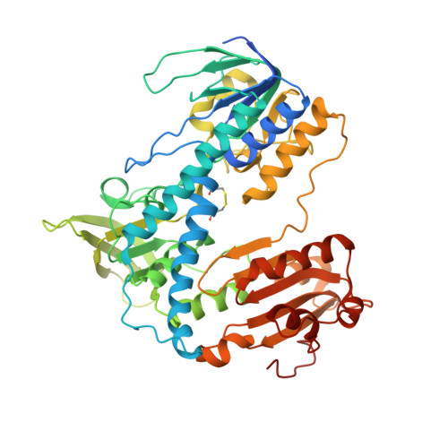
Crystal structures of oxidized and reduced mitochondrial thioredoxin reductase provide molecular details of the reaction mechanism.
Biterova, E.I., Turanov, A.A., Gladyshev, V.N., Barycki, J.J.(2005) Proc Natl Acad Sci U S A 102: 15018-15023
- PubMed: 16217027
- DOI: https://doi.org/10.1073/pnas.0504218102
- Primary Citation of Related Structures:
1ZDL, 1ZKQ - PubMed Abstract:
Thioredoxin reductase (TrxR) is an essential enzyme required for the efficient maintenance of the cellular redox homeostasis, particularly in cancer cells that are sensitive to reactive oxygen species. In mammals, distinct isozymes function in the cytosol and mitochondria. Through an intricate mechanism, these enzymes transfer reducing equivalents from NADPH to bound FAD and subsequently to an active-site disulfide. In mammalian TrxRs, the dithiol then reduces a mobile C-terminal selenocysteine-containing tetrapeptide of the opposing subunit of the dimer. Once activated, the C-terminal redox center reduces a disulfide bond within thioredoxin. In this report, we present the structural data on a mitochondrial TrxR, TrxR2 (also known as TR3 and TxnRd2). Mouse TrxR2, in which the essential selenocysteine residue had been replaced with cysteine, was isolated as a FAD-containing holoenzyme and crystallized (2.6 A; R = 22.2%; R(free) = 27.6%). The addition of NADPH to the TrxR2 crystals resulted in a color change, indicating reduction of the active-site disulfide and formation of a species presumed to be the flavin-thiolate charge transfer complex. Examination of the NADP(H)-bound model (3.0 A; R = 24.1%; R(free) = 31.2%) indicates that an active-site tyrosine residue must rotate from its initial position to stack against the nicotinamide ring of NADPH, which is juxtaposed to the isoalloxazine ring of FAD to facilitate hydride transfer. Detailed analysis of the structural data in conjunction with a model of the unusual C-terminal selenenylsulfide suggests molecular details of the reaction mechanism and highlights evolutionary adaptations among reductases.
Organizational Affiliation:
Department of Biochemistry, University of Nebraska, Lincoln, NE 68588-0664, USA.


