1Z9K

Photosynthetic Reaction Center from Rhodobacter sphaeroides


Experimental Data Snapshot

  • Method: X-RAY DIFFRACTION
  • Resolution: 4.60 Å
  • R-Value Free: 0.330 
  • R-Value Work: 0.330 
  • R-Value Observed: 0.340 

Starting Model: experimental
View more details

wwPDB Validation   3D Report Full Report


This is version 1.3 of the entry. See complete history


Literature

Design of a Redox-Linked Active Metal Site: Manganese Bound to Bacterial Reaction Centers at a Site Resembling That of Photosystem II

Uyeda, G.Camara-Artigas, A.Williams, J.C.Allen, J.P.

(2005) Biochemistry 44: 7389-7394

  • DOI: https://doi.org/10.1021/bi050377n
  • Primary Citation of Related Structures:  
    1Z9J, 1Z9K

  • PubMed Abstract: 

    Metals bound to proteins perform a number of crucial biological reactions, including the oxidation of water by a manganese cluster in photosystem II. Although evolutionarily related to photosystem II, bacterial reaction centers lack both a strong oxidant and a manganese cluster for mediating the multielectron and proton transfer needed for water oxidation. In this study, carboxylate residues were introduced by mutagenesis into highly oxidizing reaction centers at a site homologous to the manganese-binding site of photosystem II. In the presence of manganese, light-minus-dark difference optical spectra of reaction centers from the mutants showed a lack of the oxidized bacteriochlorophyll dimer, while the reduced primary quinone was still present, demonstrating that manganese was serving as a secondary electron donor. On the basis of these steady-state optical measurements, the mutant with the highest-affinity site had a dissociation constant of approximately 1 microM. For the highest-affinity mutant, a first-order rate with a lifetime of 12 ms was observed for the reduction of the oxidized bacteriochlorophyll dimer by the bound manganese upon exposure to light. The dependence of the amplitude of this component on manganese concentration yielded a dissociation constant of approximately 1 muM, similar to that observed in the steady-state measurements. The three-dimensional structure determined by X-ray diffraction of the mutant with the high-affinity site showed that the binding site contains a single bound manganese ion, three carboxylate groups (including two groups introduced by mutagenesis), a histidine residue, and a bound water molecule. These reaction centers illustrate the successful design of a redox active metal center in a protein complex.


  • Organizational Affiliation

    Department of Chemistry and Biochemistry, Arizona State University, Tempe, Arizona 85287, USA.


Macromolecules
Find similar proteins by:  (by identity cutoff)  |  3D Structure
Entity ID: 1
MoleculeChains Sequence LengthOrganismDetailsImage
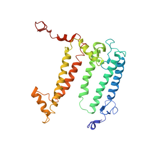
Reaction center protein L chain281Cereibacter sphaeroidesMutation(s): 0 
Gene Names: pufL
Membrane Entity: Yes 
UniProt
Find proteins for P0C0Y8 (Cereibacter sphaeroides)
Explore P0C0Y8 
Go to UniProtKB:  P0C0Y8
Entity Groups  
Sequence Clusters30% Identity50% Identity70% Identity90% Identity95% Identity100% Identity
UniProt GroupP0C0Y8
Sequence Annotations
Expand
  • Reference Sequence
Find similar proteins by:  (by identity cutoff)  |  3D Structure
Entity ID: 2
MoleculeChains Sequence LengthOrganismDetailsImage
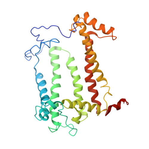
Reaction center protein M chain307Cereibacter sphaeroidesMutation(s): 0 
Gene Names: pufM
Membrane Entity: Yes 
UniProt
Find proteins for P0C0Y9 (Cereibacter sphaeroides)
Explore P0C0Y9 
Go to UniProtKB:  P0C0Y9
Entity Groups  
Sequence Clusters30% Identity50% Identity70% Identity90% Identity95% Identity100% Identity
UniProt GroupP0C0Y9
Sequence Annotations
Expand
  • Reference Sequence
Find similar proteins by:  (by identity cutoff)  |  3D Structure
Entity ID: 3
MoleculeChains Sequence LengthOrganismDetailsImage
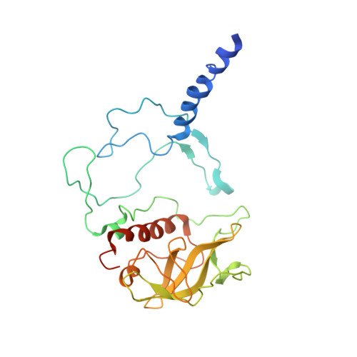
Reaction center protein H chain260Cereibacter sphaeroidesMutation(s): 0 
Gene Names: puhA
Membrane Entity: Yes 
UniProt
Find proteins for P0C0Y7 (Cereibacter sphaeroides)
Explore P0C0Y7 
Go to UniProtKB:  P0C0Y7
Entity Groups  
Sequence Clusters30% Identity50% Identity70% Identity90% Identity95% Identity100% Identity
UniProt GroupP0C0Y7
Sequence Annotations
Expand
  • Reference Sequence
Small Molecules
Ligands 5 Unique
IDChains Name / Formula / InChI Key2D Diagram3D Interactions
BCL
Query on BCL

Download Ideal Coordinates CCD File 
D [auth A],
E [auth A],
I [auth B],
J [auth B]
BACTERIOCHLOROPHYLL A
C55 H74 Mg N4 O6
DSJXIQQMORJERS-AGGZHOMASA-M
BPH
Query on BPH

Download Ideal Coordinates CCD File 
F [auth A],
K [auth B]
BACTERIOPHEOPHYTIN A
C55 H76 N4 O6
KWOZSBGNAHVCKG-SZQBJALDSA-N
U10
Query on U10

Download Ideal Coordinates CCD File 
G [auth A],
L [auth B]
UBIQUINONE-10
C59 H90 O4
ACTIUHUUMQJHFO-UPTCCGCDSA-N
FE
Query on FE

Download Ideal Coordinates CCD File 
H [auth B]FE (III) ION
Fe
VTLYFUHAOXGGBS-UHFFFAOYSA-N
MN
Query on MN

Download Ideal Coordinates CCD File 
M [auth C]MANGANESE (II) ION
Mn
WAEMQWOKJMHJLA-UHFFFAOYSA-N
Experimental Data & Validation

Experimental Data

  • Method: X-RAY DIFFRACTION
  • Resolution: 4.60 Å
  • R-Value Free: 0.330 
  • R-Value Work: 0.330 
  • R-Value Observed: 0.340 
  • Space Group: P 42 2 2
Unit Cell:
Length ( Å )Angle ( ˚ )
a = 207.8α = 90
b = 207.8β = 90
c = 107.5γ = 90
Software Package:
Software NamePurpose
MOSFLMdata reduction
SCALAdata scaling
CNSrefinement
CCP4data scaling
CNSphasing

Structure Validation

View Full Validation Report


Entry History 

Deposition Data

Revision History  (Full details and data files)

  • Version 1.0: 2005-06-07
    Type: Initial release
  • Version 1.1: 2008-04-30
    Changes: Version format compliance
  • Version 1.2: 2011-07-13
    Changes: Non-polymer description, Version format compliance
  • Version 1.3: 2023-08-23
    Changes: Data collection, Database references, Derived calculations, Refinement description