1Z0T | pdb_00001z0t

Crystal Structure of A. fulgidus Lon proteolytic domain


Experimental Data Snapshot

  • Method: X-RAY DIFFRACTION
  • Resolution: 3.00 Å
  • R-Value Free: 
    0.330 (Depositor), 0.343 (DCC) 
  • R-Value Work: 
    0.200 (Depositor), 0.211 (DCC) 
  • R-Value Observed: 
    0.206 (Depositor) 

wwPDB Validation 3D Report Full Report

Validation slider image for 1Z0T

This is version 1.4 of the entry. See complete history

Literature

Pathological crystallography: case studies of several unusual macromolecular crystals.

Dauter, Z.Botos, I.LaRonde-LeBlanc, N.Wlodawer, A.

(2005) Acta Crystallogr D Biol Crystallogr 61: 967-975

  • DOI: https://doi.org/10.1107/S0907444905011285
  • Primary Citation Related Structures: 
    1Z0T, 1Z0V

  • PubMed Abstract: 

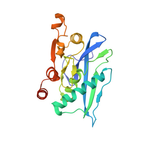
    Although macromolecular crystallography is rapidly becoming largely routine owing to advances in methods of data collection, structure solution and refinement, difficult cases are still common. To remind structural biologists about the kinds of crystallographic difficulties that might be encountered, case studies of several successfully completed structure determinations that utilized less than perfect crystals are discussed here. The structure of the proteolytic domain of Archaeoglobus fulgidus Lon was solved with crystals that contained superimposed orthorhombic and monoclinic lattices, a case not previously described for proteins. Another hexagonal crystal form of this protein exhibited an unusually high degree of non-isomorphism. Crystals of A. fulgidus Rio1 kinase exhibited both pseudosymmetry and twinning. Ways of identifying the observed phenomena and approaches to solving and refining macromolecular structures when only less than perfect crystals are available are discussed here.


  • Organizational Affiliation
    • Synchrotron Radiation Research Section, Macromolecular Crystallography Laboratory, National Cancer Institute and Biosciences Division, Argonne National Laboratory, Argonne, IL 60439, USA.

Macromolecule Content 

  • Total Structure Weight: 131.04 kDa 
  • Atom Count: 9,870 
  • Modeled Residue Count: 1,174 
  • Deposited Residue Count: 1,230 
  • Unique protein chains: 1

Macromolecules

Find similar proteins by:|  3D Structure
Entity ID: 1
MoleculeChains  Sequence LengthOrganismDetailsImage
Putative protease La homolog type
A, B, C, D, E
A, B, C, D, E, F
205Archaeoglobus fulgidusMutation(s): 0 
EC: 3.4.21.53 (PDB Primary Data), 3.4.21 (UniProt)
UniProt
Find proteins for O29883 (Archaeoglobus fulgidus (strain ATCC 49558 / DSM 4304 / JCM 9628 / NBRC 100126 / VC-16))
Explore O29883 
Go to UniProtKB:  O29883
Entity Groups
Sequence Clusters30% Identity50% Identity70% Identity90% Identity95% Identity100% Identity
UniProt GroupO29883
Sequence Annotations
Expand
Reference Sequence

Experimental Data & Validation

Experimental Data

  • Method: X-RAY DIFFRACTION
  • Resolution: 3.00 Å
  • R-Value Free:  0.330 (Depositor), 0.343 (DCC) 
  • R-Value Work:  0.200 (Depositor), 0.211 (DCC) 
  • R-Value Observed: 0.206 (Depositor) 
Space Group: P 21 21 21
Unit Cell:
Length ( Å )Angle ( ˚ )
a = 86.252α = 90
b = 90.554β = 90
c = 147.951γ = 90
Software Package:
Software NamePurpose
REFMACrefinement
MAR345data collection
SCALEPACKdata scaling
EPMRphasing

Structure Validation

View Full Validation Report


Entry History 

Deposition Data

Revision History  (Full details and data files)

  • Version 1.0: 2005-08-02
    Type: Initial release
  • Version 1.1: 2008-04-30
    Changes: Version format compliance
  • Version 1.2: 2011-07-13
    Changes: Advisory, Version format compliance
  • Version 1.3: 2017-10-11
    Changes: Refinement description
  • Version 1.4: 2024-02-14
    Changes: Data collection, Database references