1YQ9 | pdb_00001yq9

Structure of the unready oxidized form of [NiFe] hydrogenase


Experimental Data Snapshot

  • Method: X-RAY DIFFRACTION
  • Resolution: 2.35 Å
  • R-Value Free: 
    0.183 (Depositor), 0.198 (DCC) 
  • R-Value Work: 
    0.132 (Depositor), 0.150 (DCC) 
  • R-Value Observed: 
    0.135 (Depositor) 

Starting Model: experimental
View more details

wwPDB Validation 3D Report Full Report

Validation slider image for 1YQ9

This is version 1.4 of the entry. See complete history

Literature

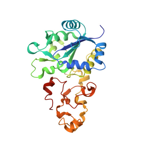
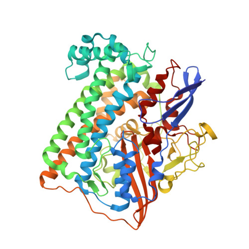
Structural differences between the ready and unready oxidized states of [NiFe] hydrogenases.

Volbeda, A.Martin, L.Cavazza, C.Matho, M.Faber, B.W.Roseboom, W.Albracht, S.P.Garcin, E.Rousset, M.Fontecilla-Camps, J.C.

(2005) J Biol Inorg Chem 10: 239-249

  • DOI: https://doi.org/10.1007/s00775-005-0632-x
  • Primary Citation Related Structures: 
    1YQ9, 1YQW, 1YRQ

  • PubMed Abstract: 

    [NiFe] hydrogenases catalyze the reversible heterolytic cleavage of molecular hydrogen. Several oxidized, inactive states of these enzymes are known that are distinguishable by their very different activation properties. So far, the structural basis for this difference has not been understood because of lack of relevant crystallographic data. Here, we present the crystal structure of the ready Ni-B state of Desulfovibrio fructosovorans [NiFe] hydrogenase and show it to have a putative mu-hydroxo Ni-Fe bridging ligand at the active site. On the other hand, a new, improved refinement procedure of the X-ray diffraction data obtained for putative unready Ni-A/Ni-SU states resulted in a more elongated electron density for the bridging ligand, suggesting that it is a diatomic species. The slow activation of the Ni-A state, compared with the rapid activation of the Ni-B state, is therefore proposed to result from the different chemical nature of the ligands in the two oxidized species. Our results along with very recent electrochemical studies suggest that the diatomic ligand could be hydro-peroxide.


  • Organizational Affiliation
    • Laboratoire de Cristallographie et de Cristallogenèse des Protèines, Institut de Biologie Structurale J.P. Ebel (CEA-CNRS-UJF), 41 rue Jules Horowitz, 38027, Grenoble Cédex 1, France. anne.volbeda@ibs.fr

Macromolecule Content 

  • Total Structure Weight: 179.3 kDa 
  • Atom Count: 12,882 
  • Modeled Residue Count: 1,581 
  • Deposited Residue Count: 1,600 
  • Unique protein chains: 2

Macromolecules

Find similar proteins by:|  3D Structure
Entity ID: 1
MoleculeChains  Sequence LengthOrganismDetailsImage
Periplasmic [NiFe] hydrogenase small subunitA,
C [auth B]
264Megalodesulfovibrio gigasMutation(s): 0 
EC: 1.12.2.1
UniProt
Find proteins for P12943 (Megalodesulfovibrio gigas)
Explore P12943 
Go to UniProtKB:  P12943
Entity Groups
Sequence Clusters30% Identity50% Identity70% Identity90% Identity95% Identity100% Identity
UniProt GroupP12943
Sequence Annotations
Expand
Reference Sequence
Find similar proteins by:|  3D Structure
Entity ID: 2
MoleculeChains  Sequence LengthOrganismDetailsImage
Periplasmic [NiFe] hydrogenase large subunitB [auth H],
D [auth I]
536Megalodesulfovibrio gigasMutation(s): 0 
EC: 1.12.2.1
UniProt
Find proteins for P12944 (Megalodesulfovibrio gigas)
Explore P12944 
Go to UniProtKB:  P12944
Entity Groups
Sequence Clusters30% Identity50% Identity70% Identity90% Identity95% Identity100% Identity
UniProt GroupP12944
Sequence Annotations
Expand
Reference Sequence

Small Molecules

Ligands 8 Unique
IDChains Name / Formula / InChI Key2D Diagram3D Interactions
SF4

Query on SF4


Download:Ideal Coordinates CCD File
E [auth A],
G [auth A],
T [auth B],
V [auth B]
IRON/SULFUR CLUSTER
Fe4 S4
LJBDFODJNLIPKO-UHFFFAOYSA-N
F3S

Query on F3S


Download:Ideal Coordinates CCD File
F [auth A],
U [auth B]
FE3-S4 CLUSTER
Fe3 S4
FCXHZBQOKRZXKS-UHFFFAOYSA-N
FCO

Query on FCO


Download:Ideal Coordinates CCD File
AA [auth I],
O [auth H]
CARBONMONOXIDE-(DICYANO) IRON
C3 Fe N2 O
VBQUCMTXYFMTTE-UHFFFAOYSA-N
GOL

Query on GOL


Download:Ideal Coordinates CCD File
I [auth A]
J [auth A]
K [auth A]
L [auth A]
R [auth H]
I [auth A],
J [auth A],
K [auth A],
L [auth A],
R [auth H],
S [auth H],
X [auth B]
GLYCEROL
C3 H8 O3
PEDCQBHIVMGVHV-UHFFFAOYSA-N
NI

Query on NI


Download:Ideal Coordinates CCD File
M [auth H],
Y [auth I]
NICKEL (II) ION
Ni
VEQPNABPJHWNSG-UHFFFAOYSA-N
H2S

Query on H2S


Download:Ideal Coordinates CCD File
CA [auth I],
H [auth A],
Q [auth H],
W [auth B]
HYDROSULFURIC ACID
H2 S
RWSOTUBLDIXVET-UHFFFAOYSA-N
PER

Query on PER


Download:Ideal Coordinates CCD File
BA [auth I],
P [auth H]
PEROXIDE ION
O2
ANAIPYUSIMHBEL-UHFFFAOYSA-N
MG

Query on MG


Download:Ideal Coordinates CCD File
N [auth H],
Z [auth I]
MAGNESIUM ION
Mg
JLVVSXFLKOJNIY-UHFFFAOYSA-N

Experimental Data & Validation

Experimental Data

  • Method: X-RAY DIFFRACTION
  • Resolution: 2.35 Å
  • R-Value Free:  0.183 (Depositor), 0.198 (DCC) 
  • R-Value Work:  0.132 (Depositor), 0.150 (DCC) 
  • R-Value Observed: 0.135 (Depositor) 
Space Group: P 1
Unit Cell:
Length ( Å )Angle ( ˚ )
a = 62.75α = 89.33
b = 93.44β = 102.41
c = 69.03γ = 90.95
Software Package:
Software NamePurpose
REFMACrefinement
PDB_EXTRACTdata extraction
ProDCdata collection
XDSdata scaling
AMoREphasing

Structure Validation

View Full Validation Report


Entry History 

Deposition Data

Revision History  (Full details and data files)

  • Version 1.0: 2005-04-19
    Type: Initial release
  • Version 1.1: 2008-04-30
    Changes: Version format compliance
  • Version 1.2: 2011-07-13
    Changes: Advisory, Refinement description, Version format compliance
  • Version 1.3: 2017-10-11
    Changes: Refinement description
  • Version 1.4: 2023-08-23
    Changes: Data collection, Database references, Derived calculations, Refinement description