1YPV | pdb_00001ypv

Structure of human thymidylate synthase at low salt conditions


Experimental Data Snapshot

  • Method: X-RAY DIFFRACTION
  • Resolution: 1.80 Å
  • R-Value Free: 
    0.243 (Depositor), 0.246 (DCC) 
  • R-Value Work: 
    0.227 (Depositor), 0.230 (DCC) 
  • R-Value Observed: 
    0.227 (Depositor) 

wwPDB Validation 3D Report Full Report

Validation slider image for 1YPV

This is version 1.3 of the entry. See complete history

Literature

Structure of human thymidylate synthase under low-salt conditions.

Lovelace, L.L.Minor, W.Lebioda, L.

(2005) Acta Crystallogr D Biol Crystallogr 61: 622-627

  • DOI: https://doi.org/10.1107/S0907444905005895
  • Primary Citation Related Structures: 
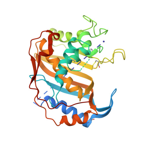
    1YPV

  • PubMed Abstract: 

    Human thymidylate synthase, a target in cancer chemotherapy, was crystallized from PEG 3350 with 30 mM ammonium sulfate (AS) in the crystallization medium. The crystals are isomorphous with the high-salt crystals ( approximately 2.0 M AS) and the structure has been solved and refined (R = 22.6%, R(free) = 24.3%) at 1.8 A resolution. The high- and low-AS-concentration structures are quite similar, with loop 181-197 is in the inactive conformation. Also, residues 95-106 and 129-135 (eukaryotic inserts region) show high mobility as assessed by poor electron density and high values of crystallographic temperature factors (residues 1-25 and 108-129 are disordered in both structures). The high mobility of this region may reflect the situation at physiological ionic strength. Of the four sulfate ions observed bound at 2.0 M AS, only two are present at 30 mM AS. The inactive conformation appears to be stabilized by the side chain of Val3 or a leucine residue from the disordered regions. The low-salt conditions of these crystals should be much more suitable for the study of thymidylate synthase inhibitors, especially those that utilize sulfate-binding sites to stabilize the inactive conformation of loop 181-197.


  • Organizational Affiliation
    • Department of Chemistry and Biochemistry, University of South Carolina, Columbia, SC 29208, USA.

Macromolecule Content 

  • Total Structure Weight: 36.23 kDa 
  • Atom Count: 2,250 
  • Modeled Residue Count: 264 
  • Deposited Residue Count: 313 
  • Unique protein chains: 1

Macromolecules

Find similar proteins by:|  3D Structure
Entity ID: 1
MoleculeChains  Sequence LengthOrganismDetailsImage
Thymidylate synthase313Homo sapiensMutation(s): 4 
Gene Names: TYMSTS
EC: 2.1.1.45
UniProt & NIH Common Fund Data Resources
Find proteins for P04818 (Homo sapiens)
Explore P04818 
Go to UniProtKB:  P04818
PHAROS:  P04818
GTEx:  ENSG00000176890 
Entity Groups
Sequence Clusters30% Identity50% Identity70% Identity90% Identity95% Identity100% Identity
UniProt GroupP04818
Sequence Annotations
Expand
Reference Sequence

Small Molecules

Modified Residues  2 Unique
IDChains TypeFormula2D DiagramParent
CME
Query on CME
A
L-PEPTIDE LINKINGC5 H11 N O3 S2CYS
SCH
Query on SCH
A
L-PEPTIDE LINKINGC4 H9 N O2 S2CYS

Experimental Data & Validation

Experimental Data

  • Method: X-RAY DIFFRACTION
  • Resolution: 1.80 Å
  • R-Value Free:  0.243 (Depositor), 0.246 (DCC) 
  • R-Value Work:  0.227 (Depositor), 0.230 (DCC) 
  • R-Value Observed: 0.227 (Depositor) 
Space Group: P 31 2 1
Unit Cell:
Length ( Å )Angle ( ˚ )
a = 95.772α = 90
b = 95.772β = 90
c = 81.851γ = 120
Software Package:
Software NamePurpose
d*TREKdata scaling
SCALEPACKdata scaling
CNSrefinement
HKL-2000data reduction
CNSphasing

Structure Validation

View Full Validation Report


Entry History 

Deposition Data

Revision History  (Full details and data files)

  • Version 1.0: 2005-04-26
    Type: Initial release
  • Version 1.1: 2008-04-30
    Changes: Version format compliance
  • Version 1.2: 2011-07-13
    Changes: Derived calculations, Version format compliance
  • Version 1.3: 2022-04-13
    Changes: Database references, Derived calculations, Structure summary