1YN5 | pdb_00001yn5

Crystal Structures of EAP Domains from Staphylococcus aureus Reveal an Unexpected Homology to Bacterial Superantigens


Experimental Data Snapshot

  • Method: X-RAY DIFFRACTION
  • Resolution: 2.20 Å
  • R-Value Free: 
    0.317 (Depositor), 0.314 (DCC) 
  • R-Value Work: 
    0.258 (Depositor), 0.256 (DCC) 
  • R-Value Observed: 
    0.258 (Depositor) 

Starting Model: experimental
View more details

wwPDB Validation 3D Report Full Report

Validation slider image for 1YN5

This is version 1.3 of the entry. See complete history

Literature

The Crystal Structures of EAP Domains from Staphylococcus aureus Reveal an Unexpected Homology to Bacterial Superantigens.

Geisbrecht, B.V.Hamaoka, B.Y.Perman, B.Zemla, A.Leahy, D.J.

(2005) J Biological Chem 280: 17243-17250

  • DOI: https://doi.org/10.1074/jbc.M412311200
  • Primary Citation Related Structures: 
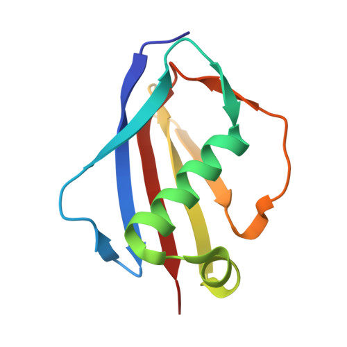
    1YN3, 1YN4, 1YN5

  • PubMed Abstract: 

    The Eap (extracellular adherence protein) of Staphylococcus aureus functions as a secreted virulence factor by mediating interactions between the bacterial cell surface and several extracellular host proteins. Eap proteins from different Staphylococcal strains consist of four to six tandem repeats of a structurally uncharacterized domain (EAP domain). We have determined the three-dimensional structures of three different EAP domains to 1.8, 2.2, and 1.35 A resolution, respectively. These structures reveal a core fold that is comprised of an alpha-helix lying diagonally across a five-stranded, mixed beta-sheet. Comparison of EAP domains with known structures reveals an unexpected homology with the C-terminal domain of bacterial superantigens. Examination of the structure of the superantigen SEC2 bound to the beta-chain of a T-cell receptor suggests a possible ligand-binding site within the EAP domain (Fields, B. A., Malchiodi, E. L., Li, H., Ysern, X., Stauffacher, C. V., Schlievert, P. M., Karjalainen, K., and Mariuzza, R. (1996) Nature 384, 188-192). These results provide the first structural characterization of EAP domains, relate EAP domains to a large class of bacterial toxins, and will guide the design of future experiments to analyze EAP domain structure/function relationships.


  • Organizational Affiliation
    • Department of Biophysics and Biophysical Chemistry, The Johns Hopkins University School of Medicine, Baltimore, MD 21205, USA.

Macromolecule Content 

  • Total Structure Weight: 23.33 kDa 
  • Atom Count: 1,667 
  • Modeled Residue Count: 203 
  • Deposited Residue Count: 206 
  • Unique protein chains: 1

Macromolecules

Find similar proteins by:|  3D Structure
Entity ID: 1
MoleculeChains  Sequence LengthOrganismDetailsImage
EapH2
A, B
103Staphylococcus aureusMutation(s): 0 
UniProt
Find proteins for A0A0H3JUK5 (Staphylococcus aureus (strain Mu50 / ATCC 700699))
Explore A0A0H3JUK5 
Go to UniProtKB:  A0A0H3JUK5
Entity Groups
Sequence Clusters30% Identity50% Identity70% Identity90% Identity95% Identity100% Identity
UniProt GroupA0A0H3JUK5
Sequence Annotations
Expand
Reference Sequence

Experimental Data & Validation

Experimental Data

  • Method: X-RAY DIFFRACTION
  • Resolution: 2.20 Å
  • R-Value Free:  0.317 (Depositor), 0.314 (DCC) 
  • R-Value Work:  0.258 (Depositor), 0.256 (DCC) 
  • R-Value Observed: 0.258 (Depositor) 
Space Group: P 1 21 1
Unit Cell:
Length ( Å )Angle ( ˚ )
a = 50.01α = 90
b = 31.97β = 105.21
c = 67.98γ = 90
Software Package:
Software NamePurpose
DENZOdata reduction
SCALEPACKdata scaling
MOLREPphasing
CNSrefinement

Structure Validation

View Full Validation Report


Entry History 

Deposition Data

Revision History  (Full details and data files)

  • Version 1.0: 2005-03-01
    Type: Initial release
  • Version 1.1: 2008-04-30
    Changes: Version format compliance
  • Version 1.2: 2011-07-13
    Changes: Version format compliance
  • Version 1.3: 2023-08-23
    Changes: Data collection, Database references, Refinement description