1YMG

The Channel Architecture of Aquaporin O at 2.2 Angstrom Resolution


Experimental Data Snapshot

  • Method: X-RAY DIFFRACTION
  • Resolution: 2.24 Å
  • R-Value Free: 0.303 
  • R-Value Work: 0.240 
  • R-Value Observed: 0.240 

Starting Model: experimental
View more details

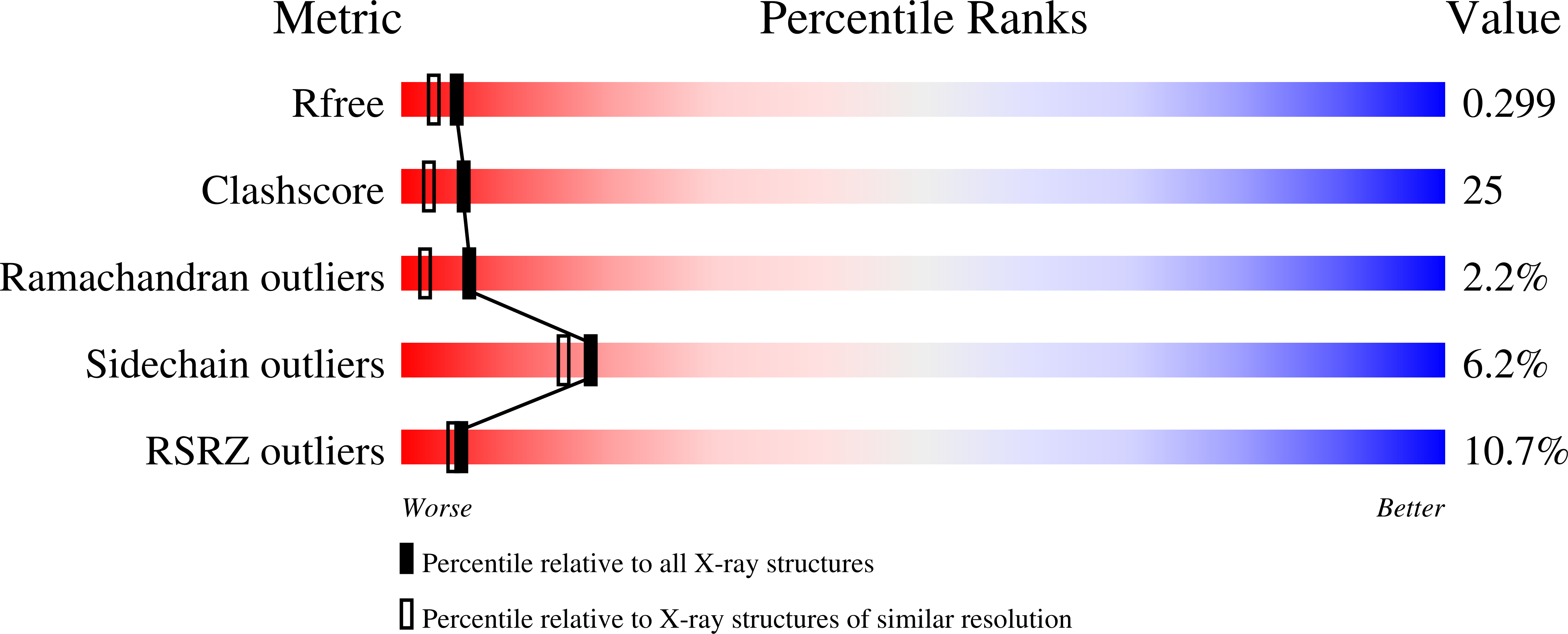
wwPDB Validation   3D Report Full Report


Ligand Structure Quality Assessment 


This is version 2.1 of the entry. See complete history

Re-refinement Note

A newer entry is available that reflects an alternative modeling of the original data: 2B6P


Literature

The Channel Architecture of Aquaporin 0 at a 2.2-A Resolution

Harries, W.E.C.Akhavan, D.Miercke, L.J.W.Khademi, S.Stroud, R.M.

(2004) Proc Natl Acad Sci U S A 101: 14045-14050

  • DOI: https://doi.org/10.1073/pnas.0405274101
  • Primary Citation of Related Structures:  
    1YMG

  • PubMed Abstract: 

    We determined the x-ray structure of bovine aquaporin 0 (AQP0) to a resolution of 2.2 A. The structure of this eukaryotic, integral membrane protein suggests that the selectivity of AQP0 for water transport is based on the identity and location of signature amino acid residues that are hallmarks of the water-selective arm of the AQP family of proteins. Furthermore, the channel lumen is narrowed only by two, quasi-2-fold related tyrosine side chains that might account for reduced water conductance relative to other AQPs. The channel is functionally open to the passage of water because there are eight discreet water molecules within the channel. Comparison of this structure with the recent electron-diffraction structure of the junctional form of sheep AQP0 at pH 6.0 that was interpreted as closed shows no global change in the structure of AQP0 and only small changes in side-chain positions. We observed no structural change to the channel or the molecule as a whole at pH 10, which could be interpreted as the postulated pH-gating mechanism of AQP0-mediated water transport at pH >6.5. Contrary to the electron-diffraction structure, the comparison shows no evidence of channel gating induced by association of the extracellular domains of AQP0 at pH 6.0. Our structure aids the analysis of the interaction of the extracellular domains and the possibility of a cell-cell adhesion role for AQP0. In addition, our structure illustrates the basis for formation of certain types of cataracts that are the result of mutations.


  • Organizational Affiliation

    Macromolecular Structure Group, Department of Biochemistry and Biophysics, University of California, S-412C Genentech Hall, 600 16th Street, San Francisco, CA 94143-2240, USA.


Macromolecules
Find similar proteins by:  (by identity cutoff)  |  3D Structure
Entity ID: 1
MoleculeChains Sequence LengthOrganismDetailsImage
Lens fiber major intrinsic protein263Bos taurusMutation(s): 0 
Membrane Entity: Yes 
UniProt
Find proteins for P06624 (Bos taurus)
Explore P06624 
Go to UniProtKB:  P06624
Entity Groups  
Sequence Clusters30% Identity50% Identity70% Identity90% Identity95% Identity100% Identity
UniProt GroupP06624
Sequence Annotations
Expand
  • Reference Sequence
Small Molecules
Ligands 1 Unique
IDChains Name / Formula / InChI Key2D Diagram3D Interactions
BNG
Query on BNG

Download Ideal Coordinates CCD File 
B [auth A],
C [auth A]
nonyl beta-D-glucopyranoside
C15 H30 O6
QFAPUKLCALRPLH-UXXRCYHCSA-N
Experimental Data & Validation

Experimental Data

  • Method: X-RAY DIFFRACTION
  • Resolution: 2.24 Å
  • R-Value Free: 0.303 
  • R-Value Work: 0.240 
  • R-Value Observed: 0.240 
  • Space Group: P 4 21 2
Unit Cell:
Length ( Å )Angle ( ˚ )
a = 110.531α = 90
b = 110.531β = 90
c = 53.39γ = 90
Software Package:
Software NamePurpose
DENZOdata reduction
SCALEPACKdata scaling
CNSrefinement
CNSphasing

Structure Validation

View Full Validation Report


Ligand Structure Quality Assessment 


Entry History 

Deposition Data

Revision History  (Full details and data files)

  • Version 1.0: 2005-02-08
    Type: Initial release
  • Version 1.1: 2008-04-30
    Changes: Version format compliance
  • Version 1.2: 2011-07-13
    Changes: Derived calculations, Version format compliance
  • Version 1.3: 2012-05-09
    Changes: Structure summary
  • Version 1.4: 2012-06-20
    Changes: Structure summary
  • Version 2.0: 2020-07-29
    Type: Remediation
    Reason: Carbohydrate remediation
    Changes: Atomic model, Data collection, Derived calculations, Structure summary
  • Version 2.1: 2023-08-23
    Changes: Data collection, Database references, Refinement description, Structure summary