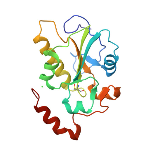
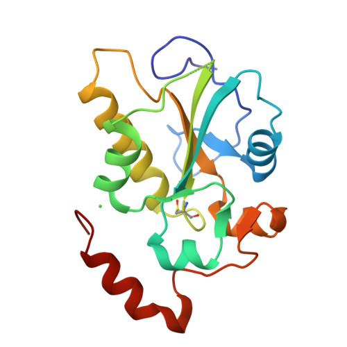
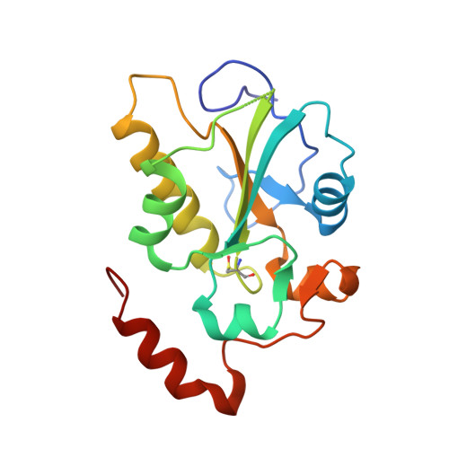
1YM9

Crystal structure of the CDC25B phosphatase catalytic domain with the active site cysteine in the sulfinic form


Experimental Data Snapshot

  • Method: X-RAY DIFFRACTION
  • Resolution: 2.00 Å
  • R-Value Free: 0.237 
  • R-Value Work: 0.218 
  • R-Value Observed: 0.218 

Starting Model: experimental
View more details

wwPDB Validation   3D Report Full Report


This is version 1.6 of the entry. See complete history


Literature

Structural Mechanism of Oxidative Regulation of the Phosphatase Cdc25B via an Intramolecular Disulfide Bond

Buhrman, G.K.Parker, B.Sohn, J.Rudolph, J.Mattos, C.

(2005) Biochemistry 44: 5307-5316

  • DOI: https://doi.org/10.1021/bi047449f
  • Primary Citation of Related Structures:  
    1YM9, 1YMD, 1YMK, 1YML, 1YS0

  • PubMed Abstract: 

    Cdc25B phosphatase, an important regulator of the cell cycle, forms an intramolecular disulfide bond in response to oxidation leading to reversible inactivation of phosphatase activity. We have obtained a crystallographic time course revealing the structural rearrangements that occur in the P-loop as the enzyme goes from its apo state, through the sulfenic (Cys-SO(-)) intermediate, to the stable disulfide. We have also obtained the structures of the irreversibly oxidized sulfinic (Cys-SO(2)(-)) and sulfonic (Cys-SO(3)(-)) Cdc25B. The active site P-loop is found in three conformations. In the apoenzyme, the P-loop is in the active conformation. In the sulfenic intermediate, the P-loop partially obstructs the active site cysteine, poised to undergo the conformational changes that accompany disulfide bond formation. In the disulfide form, the P-loop is closed over the active site cysteine, resulting in an enzyme that is unable to bind substrate. The structural changes that occur in the sulfenic intermediate of Cdc25B are distinctly different from those seen in protein tyrosine phosphatase 1B where a five-membered sulfenyl amide ring is generated as the stable end product. This work elucidates the mechanism by which chemistry and structure are coupled in the regulation of Cdc25B by reactive oxygen species.


  • Organizational Affiliation

    Department of Molecular and Structural Biochemistry, 128 Polk Hall, CB 7622, North Carolina State University, Raleigh, North Carolina 27695, USA.


Macromolecules
Find similar proteins by:  (by identity cutoff)  |  3D Structure
Entity ID: 1
MoleculeChains Sequence LengthOrganismDetailsImage
M-phase inducer phosphatase 2175Homo sapiensMutation(s): 1 
Gene Names: CDC25BCDC25HU2
EC: 3.1.3.48
UniProt & NIH Common Fund Data Resources
Find proteins for P30305 (Homo sapiens)
Explore P30305 
Go to UniProtKB:  P30305
PHAROS:  P30305
GTEx:  ENSG00000101224 
Entity Groups  
Sequence Clusters30% Identity50% Identity70% Identity90% Identity95% Identity100% Identity
UniProt GroupP30305
Sequence Annotations
Expand
  • Reference Sequence
Small Molecules
Ligands 1 Unique
IDChains Name / Formula / InChI Key2D Diagram3D Interactions
CL
Query on CL

Download Ideal Coordinates CCD File 
B [auth A]CHLORIDE ION
Cl
VEXZGXHMUGYJMC-UHFFFAOYSA-M
Modified Residues  1 Unique
IDChains TypeFormula2D DiagramParent
CSD
Query on CSD
A
L-PEPTIDE LINKINGC3 H7 N O4 SCYS
Experimental Data & Validation

Experimental Data

  • Method: X-RAY DIFFRACTION
  • Resolution: 2.00 Å
  • R-Value Free: 0.237 
  • R-Value Work: 0.218 
  • R-Value Observed: 0.218 
  • Space Group: P 21 21 21
Unit Cell:
Length ( Å )Angle ( ˚ )
a = 49.268α = 90
b = 70.627β = 90
c = 73.761γ = 90
Software Package:
Software NamePurpose
HKL-2000data collection
SCALEPACKdata scaling
CNSrefinement
HKL-2000data reduction
CNSphasing

Structure Validation

View Full Validation Report


Entry History 

Deposition Data

Revision History  (Full details and data files)

  • Version 1.0: 2005-04-12
    Type: Initial release
  • Version 1.1: 2008-04-30
    Changes: Version format compliance
  • Version 1.2: 2011-07-13
    Changes: Version format compliance
  • Version 1.3: 2014-11-12
    Changes: Non-polymer description
  • Version 1.4: 2018-01-31
    Changes: Experimental preparation
  • Version 1.5: 2023-08-23
    Changes: Data collection, Database references, Derived calculations, Refinement description
  • Version 1.6: 2024-11-20
    Changes: Structure summary