1YLC | pdb_00001ylc

Trypsin/BPTI complex mutant


Experimental Data Snapshot

  • Method: X-RAY DIFFRACTION
  • Resolution: 1.70 Å
  • R-Value Free: 
    0.224 (Depositor), 0.224 (DCC) 
  • R-Value Work: 
    0.197 (Depositor), 0.196 (DCC) 
  • R-Value Observed: 
    0.197 (Depositor) 

wwPDB Validation 3D Report Full Report

Validation slider image for 1YLC

This is version 1.4 of the entry. See complete history

Literature

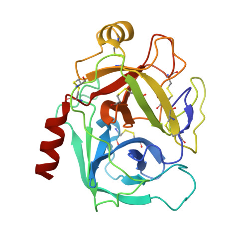
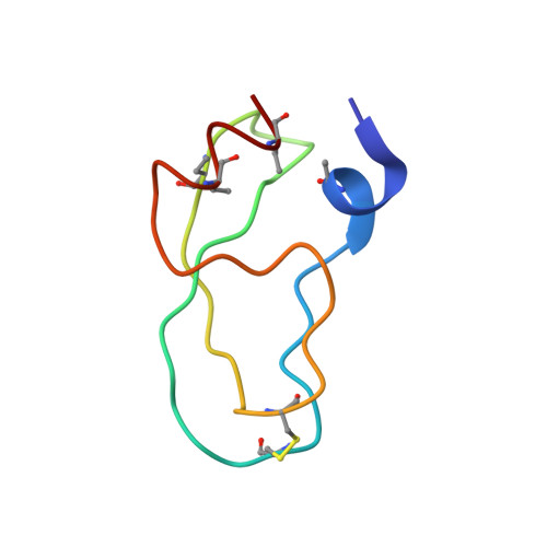
Partially folded bovine pancreatic trypsin inhibitor analogues attain fully native structures when co-crystallized with S195A rat trypsin

Getun, I.V.Brown, C.K.Tulla-Puche, J.Ohlendorf, D.Woodward, C.Barany, G.

(2008) J Mol Biology 375: 812-823

  • DOI: https://doi.org/10.1016/j.jmb.2007.10.084
  • Primary Citation Related Structures: 
    1YKT, 1YLC, 1YLD

  • PubMed Abstract: 

    Crystal structures, at 1.7 A resolution, were solved for complexes between each of two chemically synthesized partially folded analogues of bovine pancreatic trypsin inhibitor (BPTI) with the proteolytically inactive rat trypsin mutant S195A. The BPTI analogue termed [14-38](Abu) retains only the disulfide bond between Cys14 and Cys38, while Cys5, Cys30, Cys51, and Cys55 are replaced by isosteric alpha-amino-n-butyric acid residues. The analogue K26P,A27D[14-38](Abu) contains two further replacements, by statistically favored residues, in the type I beta-turn that has been suggested to be a main site for initiation of BPTI folding. As a control, the structure of the complex between S195A trypsin and wild-type BPTI was also solved. Despite significant differences in the degree of structure detected among these three BPTIs in solution by several biophysical techniques, their tertiary folds once bound to S195A trypsin in a crystalline lattice are essentially superimposable.


  • Organizational Affiliation
    • Department of Chemistry, University of Minnesota, Minneapolis, MN 55455, USA.

Macromolecule Content 

  • Total Structure Weight: 30.36 kDa 
  • Atom Count: 2,348 
  • Modeled Residue Count: 279 
  • Deposited Residue Count: 279 
  • Unique protein chains: 2

Macromolecules

Find similar proteins by:|  3D Structure
Entity ID: 1
MoleculeChains  Sequence LengthOrganismDetailsImage
Trypsin II223Rattus norvegicusMutation(s): 1 
Gene Names: Try2
EC: 3.4.21.4
UniProt
Find proteins for P00763 (Rattus norvegicus)
Explore P00763 
Go to UniProtKB:  P00763
Entity Groups
Sequence Clusters30% Identity50% Identity70% Identity90% Identity95% Identity100% Identity
UniProt GroupP00763
Sequence Annotations
Expand
Reference Sequence
Find similar proteins by:|  3D Structure
Entity ID: 2
MoleculeChains  Sequence LengthOrganismDetailsImage
Pancreatic trypsin inhibitor56N/AMutation(s): 4 
UniProt
Find proteins for P00974 (Bos taurus)
Explore P00974 
Go to UniProtKB:  P00974
Entity Groups
Sequence Clusters30% Identity50% Identity70% Identity90% Identity95% Identity100% Identity
UniProt GroupP00974
Sequence Annotations
Expand
Reference Sequence

Experimental Data & Validation

Experimental Data

  • Method: X-RAY DIFFRACTION
  • Resolution: 1.70 Å
  • R-Value Free:  0.224 (Depositor), 0.224 (DCC) 
  • R-Value Work:  0.197 (Depositor), 0.196 (DCC) 
  • R-Value Observed: 0.197 (Depositor) 
Space Group: P 32 2 1
Unit Cell:
Length ( Å )Angle ( ˚ )
a = 92.791α = 90
b = 92.791β = 90
c = 62.197γ = 120
Software Package:
Software NamePurpose
CNSrefinement
CNSphasing

Structure Validation

View Full Validation Report


Entry History 

Deposition Data

Revision History  (Full details and data files)

  • Version 1.0: 2006-04-25
    Type: Initial release
  • Version 1.1: 2008-04-30
    Changes: Version format compliance
  • Version 1.2: 2011-07-13
    Changes: Version format compliance
  • Version 1.3: 2017-10-11
    Changes: Refinement description
  • Version 1.4: 2021-10-20
    Changes: Database references, Derived calculations