1YG2 | pdb_00001yg2

Structure of the Vibrio cholerae virulence activator AphA


Experimental Data Snapshot

  • Method: X-RAY DIFFRACTION
  • Resolution: 2.20 Å
  • R-Value Free: 
    0.280 (Depositor), 0.279 (DCC) 
  • R-Value Work: 
    0.233 (Depositor), 0.234 (DCC) 
  • R-Value Observed: 
    0.233 (Depositor) 

wwPDB Validation 3D Report Full Report

Validation slider image for 1YG2

This is version 1.4 of the entry. See complete history

Literature

Crystal structure of the virulence gene activator AphA from Vibrio cholerae reveals it is a novel member of the winged helix transcription factor superfamily

De Silva, R.S.Kovacikova, G.Lin, W.Taylor, R.K.Skorupski, K.Kull, F.J.

(2005) J Biological Chem 280: 13779-13783

  • DOI: https://doi.org/10.1074/jbc.M413781200
  • Primary Citation Related Structures: 
    1YG2

  • PubMed Abstract: 

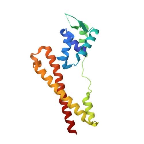
    AphA is a member of a new and largely uncharacterized family of transcriptional activators that is required for initiating virulence gene expression in Vibrio cholerae, the causative agent of the frequently fatal epidemic diarrheal disease cholera. AphA activates transcription by an unusual mechanism that appears to involve a direct interaction with the LysR-type regulator AphB at the tcpPH promoter. As a first step toward understanding the molecular basis for tcpPH activation by AphA and AphB, we have determined the crystal structure of AphA to 2.2 angstrom resolution. AphA is a dimer with an N-terminal winged helix DNA binding domain that is architecturally similar to that of the MarR family of transcriptional regulators. Unlike this family, however, AphA has a unique C-terminal antiparallel coiled coil domain that serves as its primary dimerization interface. AphA monomers are highly unstable by themselves and form a linked topology, requiring the protein to partially unfold to form the dimer. The structure of AphA also provides insights into how it cooperates with AphB to activate transcription, most likely by forming a heterotetrameric complex at the tcpPH promoter.


  • Organizational Affiliation
    • Department of Chemistry, Dartmouth College, Dartmouth Medical School, Hanover, New Hampshire 03755, USA.

Macromolecule Content 

  • Total Structure Weight: 20.45 kDa 
  • Atom Count: 1,436 
  • Modeled Residue Count: 169 
  • Deposited Residue Count: 179 
  • Unique protein chains: 1

Macromolecules

Find similar proteins by:|  3D Structure
Entity ID: 1
MoleculeChains  Sequence LengthOrganismDetailsImage
gene activator AphA179Vibrio choleraeMutation(s): 0 
Gene Names: AphA
UniProt
Find proteins for Q9X399 (Vibrio cholerae)
Explore Q9X399 
Go to UniProtKB:  Q9X399
Entity Groups
Sequence Clusters30% Identity50% Identity70% Identity90% Identity95% Identity100% Identity
UniProt GroupQ9X399
Sequence Annotations
Expand
Reference Sequence

Experimental Data & Validation

Experimental Data

  • Method: X-RAY DIFFRACTION
  • Resolution: 2.20 Å
  • R-Value Free:  0.280 (Depositor), 0.279 (DCC) 
  • R-Value Work:  0.233 (Depositor), 0.234 (DCC) 
  • R-Value Observed: 0.233 (Depositor) 
Space Group: C 2 2 21
Unit Cell:
Length ( Å )Angle ( ˚ )
a = 77.5α = 90
b = 94.65β = 90
c = 59.64γ = 90
Software Package:
Software NamePurpose
ADSCdata collection
XDSdata reduction
CNSrefinement
XDSdata scaling
CNSphasing

Structure Validation

View Full Validation Report


Entry History 

Deposition Data

Revision History  (Full details and data files)

  • Version 1.0: 2005-01-11
    Type: Initial release
  • Version 1.1: 2008-04-30
    Changes: Version format compliance
  • Version 1.2: 2011-07-13
    Changes: Derived calculations, Version format compliance
  • Version 1.3: 2018-01-31
    Changes: Experimental preparation
  • Version 1.4: 2024-02-14
    Changes: Data collection, Database references