1YCE | pdb_00001yce

Structure of the rotor ring of F-type Na+-ATPase from Ilyobacter tartaricus


Experimental Data Snapshot

  • Method: X-RAY DIFFRACTION
  • Resolution: 2.40 Å
  • R-Value Free: 
    0.246 (Depositor), 0.250 (DCC) 
  • R-Value Work: 
    0.201 (Depositor), 0.210 (DCC) 
  • R-Value Observed: 
    0.203 (Depositor) 

wwPDB Validation   3D Report Full Report


This is version 1.3 of the entry. See complete history


Literature

Structure of the rotor ring of F-Type Na+-ATPase from Ilyobacter tartaricus.

Meier, T.Polzer, P.Diederichs, K.Welte, W.Dimroth, P.

(2005) Science 308: 659-662

  • DOI: https://doi.org/10.1126/science.1111199
  • Primary Citation of Related Structures:  
    1YCE

  • PubMed Abstract: 

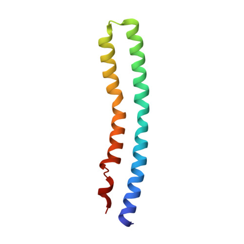
    In the crystal structure of the membrane-embedded rotor ring of the sodium ion-translocating adenosine 5'-triphosphate (ATP) synthase of Ilyobacter tartaricus at 2.4 angstrom resolution, 11 c subunits are assembled into an hourglass-shaped cylinder with 11-fold symmetry. Sodium ions are bound in a locked conformation close to the outer surface of the cylinder near the middle of the membrane. The structure supports an ion-translocation mechanism in the intact ATP synthase in which the binding site converts from the locked conformation into one that opens toward subunit a as the rotor ring moves through the subunit a/c interface.


  • Organizational Affiliation

    Institut für Mikrobiologie, Eidgenössische Technische Hochschule (ETH), Zürich Hönggerberg, Wolfgang-Pauli-Str. 10, CH-8093 Zürich, Switzerland.


Macromolecules
Find similar proteins by:  (by identity cutoff)  |  3D Structure
Entity ID: 1
MoleculeChains Sequence LengthOrganismDetailsImage
subunit c89Ilyobacter tartaricusMutation(s): 0 
Membrane Entity: Yes 
UniProt
Find proteins for Q8KRV3 (Ilyobacter tartaricus)
Explore Q8KRV3 
Go to UniProtKB:  Q8KRV3
Entity Groups  
Sequence Clusters30% Identity50% Identity70% Identity90% Identity95% Identity100% Identity
UniProt GroupQ8KRV3
Sequence Annotations
Expand
  • Reference Sequence
Small Molecules
Ligands 2 Unique
IDChains Name / Formula / InChI Key2D Diagram3D Interactions
F09
Query on F09

Download Ideal Coordinates CCD File 
BB [auth E]
BC [auth R]
BD [auth i]
BE [auth v]
DB [auth F]
NONAN-1-OL
C9 H20 O
ZWRUINPWMLAQRD-UHFFFAOYSA-N
NA
Query on NA

Download Ideal Coordinates CCD File 
AB [auth E]
AC [auth R]
AD [auth i]
AE [auth v]
CB [auth F]
SODIUM ION
Na
FKNQFGJONOIPTF-UHFFFAOYSA-N
Experimental Data & Validation

Experimental Data

  • Method: X-RAY DIFFRACTION
  • Resolution: 2.40 Å
  • R-Value Free:  0.246 (Depositor), 0.250 (DCC) 
  • R-Value Work:  0.201 (Depositor), 0.210 (DCC) 
  • R-Value Observed: 0.203 (Depositor) 
Space Group: P 1 21 1
Unit Cell:
Length ( Å )Angle ( ˚ )
a = 147.7α = 90
b = 140β = 118.4
c = 153γ = 90
Software Package:
Software NamePurpose
REFMACrefinement
XDSdata scaling
PHASERphasing

Structure Validation

View Full Validation Report


Entry History 

Deposition Data

Revision History  (Full details and data files)

  • Version 1.0: 2005-04-12
    Type: Initial release
  • Version 1.1: 2008-04-30
    Changes: Version format compliance
  • Version 1.2: 2011-07-13
    Changes: Advisory, Version format compliance
  • Version 1.3: 2024-02-14
    Changes: Data collection, Database references, Derived calculations, Refinement description