1Y8I | pdb_00001y8i

Horse methemoglobin low salt, PH 7.0 (98% relative humidity)


Experimental Data Snapshot

  • Method: X-RAY DIFFRACTION
  • Resolution: 2.60 Å
  • R-Value Free: 
    0.228 (Depositor), 0.225 (DCC) 
  • R-Value Work: 
    0.167 (Depositor), 0.164 (DCC) 
  • R-Value Observed: 
    0.167 (Depositor) 

Starting Model: experimental
View more details

wwPDB Validation 3D Report Full Report

Validation slider image for 1Y8I

Ligand Structure Quality Assessment 


This is version 1.3 of the entry. See complete history

Literature

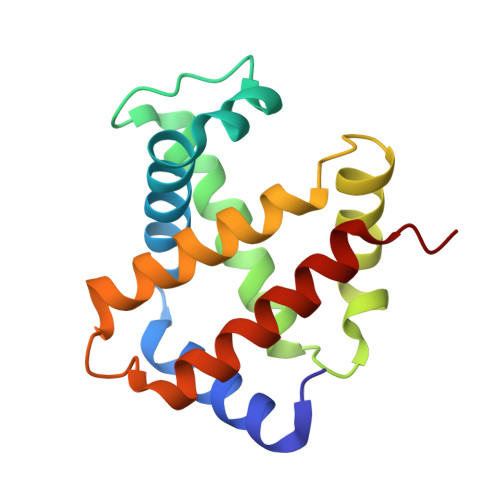
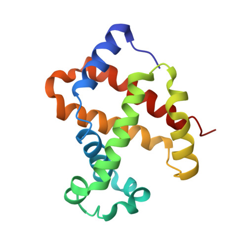
A new relaxed state in horse methemoglobin characterized by crystallographic studies.

Sankaranarayanan, R.Biswal, B.K.Vijayan, M.

(2005) Proteins 60: 547-551

  • DOI: https://doi.org/10.1002/prot.20510
  • Primary Citation Related Structures: 
    1Y8H, 1Y8I, 1Y8K

  • PubMed Abstract: 

    A new relaxed state has been characterized in the crystals of horse methemoglobin grown at neutral pH at low ionic concentration and their low humidity variants. The crystals provide an example for improvement in X-ray diffraction quality with reduced solvent content. Only the classical R state has been so far observed in liganded horse hemoglobin. The state characterized in the present study lies in between the R state and the R2 state characterized earlier in liganded human hemoglobin. The results presented here, along with those of earlier studies, suggest that relaxed and tense hemoglobin can access ensembles of states.


  • Organizational Affiliation
    • Molecular Biophysics Unit, Indian Institute of Science, Bangalore, India.

Macromolecule Content 

  • Total Structure Weight: 64.81 kDa 
  • Atom Count: 4,882 
  • Modeled Residue Count: 574 
  • Deposited Residue Count: 574 
  • Unique protein chains: 2

Macromolecules

Find similar proteins by:|  3D Structure
Entity ID: 1
MoleculeChains  Sequence LengthOrganismDetailsImage
Hemoglobin alpha chains
A, C
141Equus caballusMutation(s): 0 
UniProt
Find proteins for P01958 (Equus caballus)
Explore P01958 
Go to UniProtKB:  P01958
Entity Groups
Sequence Clusters30% Identity50% Identity70% Identity90% Identity95% Identity100% Identity
UniProt GroupP01958
Sequence Annotations
Expand
Reference Sequence
Find similar proteins by:|  3D Structure
Entity ID: 2
MoleculeChains  Sequence LengthOrganismDetailsImage
Hemoglobin beta chain
B, D
146Equus caballusMutation(s): 0 
UniProt
Find proteins for P02062 (Equus caballus)
Explore P02062 
Go to UniProtKB:  P02062
Entity Groups
Sequence Clusters30% Identity50% Identity70% Identity90% Identity95% Identity100% Identity
UniProt GroupP02062
Sequence Annotations
Expand
Reference Sequence

Experimental Data & Validation

Experimental Data

  • Method: X-RAY DIFFRACTION
  • Resolution: 2.60 Å
  • R-Value Free:  0.228 (Depositor), 0.225 (DCC) 
  • R-Value Work:  0.167 (Depositor), 0.164 (DCC) 
  • R-Value Observed: 0.167 (Depositor) 
Space Group: P 21 21 21
Unit Cell:
Length ( Å )Angle ( ˚ )
a = 62.798α = 90
b = 80.907β = 90
c = 112.159γ = 90
Software Package:
Software NamePurpose
DENZOdata reduction
SCALEPACKdata scaling
AMoREphasing
CNSrefinement

Structure Validation

View Full Validation Report


Ligand Structure Quality Assessment 


Entry History 

Deposition Data

Revision History  (Full details and data files)

  • Version 1.0: 2005-07-26
    Type: Initial release
  • Version 1.1: 2008-04-30
    Changes: Version format compliance
  • Version 1.2: 2011-07-13
    Changes: Version format compliance
  • Version 1.3: 2023-08-23
    Changes: Data collection, Database references, Derived calculations, Refinement description