1Y4J

Crystal structure of the paralogue of the human formylglycine generating enzyme


Experimental Data Snapshot

  • Method: X-RAY DIFFRACTION
  • Resolution: 1.86 Å
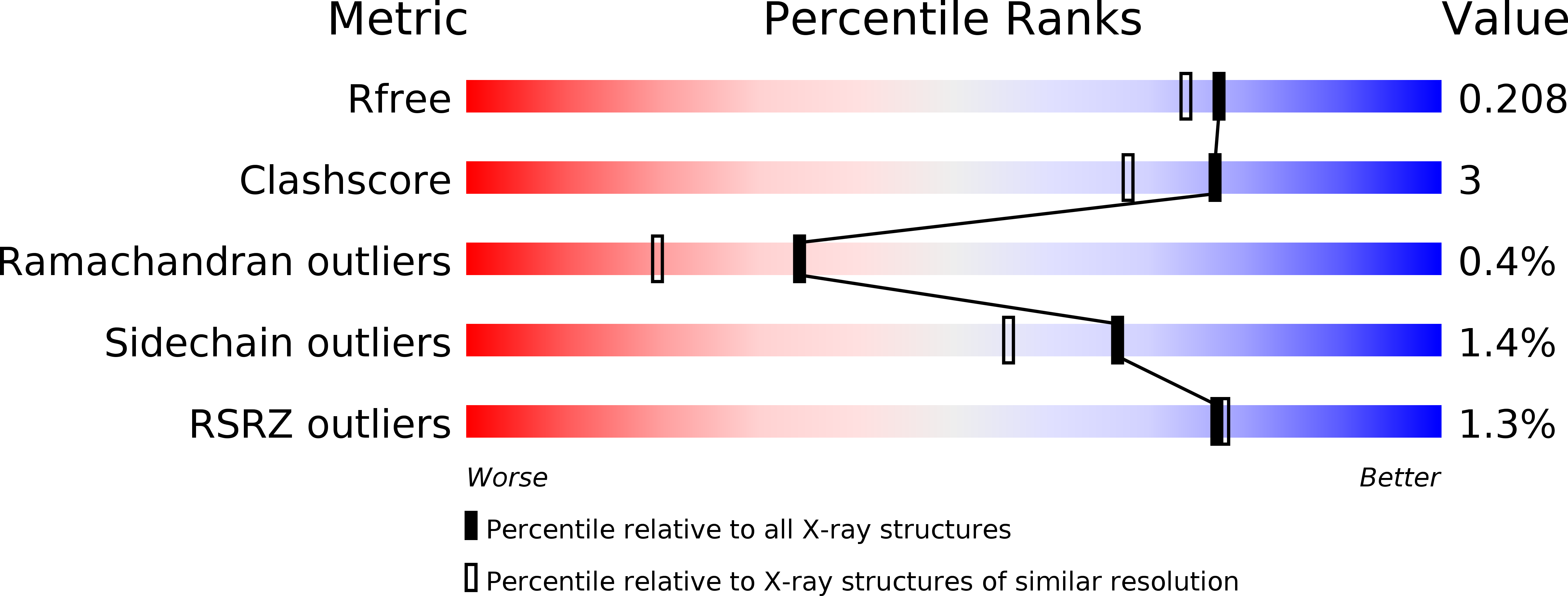
  • R-Value Free: 0.205 
  • R-Value Work: 0.170 
  • R-Value Observed: 0.172 

wwPDB Validation   3D Report Full Report


This is version 2.1 of the entry. See complete history


Literature

Crystal Structure of Human pFGE, the Paralog of the C{alpha}-formylglycine-generating Enzyme

Dickmanns, A.Schmidt, B.Rudolph, M.G.Mariappan, M.Dierks, T.von Figura, K.Ficner, R.

(2005) J Biol Chem 280: 15180-15187

  • DOI: https://doi.org/10.1074/jbc.M414317200
  • Primary Citation of Related Structures:  
    1Y4J

  • PubMed Abstract: 

    In eukaryotes, sulfate esters are degraded by sulfatases, which possess a unique Calpha-formylglycine residue in their active site. The defect in post-translational formation of the Calpha-formylglycine residue causes a severe lysosomal storage disorder in humans. Recently, FGE (formylglycine-generating enzyme) has been identified as the protein required for this specific modification. Using sequence comparisons, a protein homologous to FGE was found and denoted pFGE (paralog of FGE). pFGE binds a sulfatase-derived peptide bearing the FGE recognition motif, but it lacks formylglycine-generating activity. Both proteins belong to a large family of pro- and eukaryotic proteins containing the DUF323 domain, a formylglycine-generating enzyme domain of unknown three-dimensional structure. We have crystallized the glycosylated human pFGE and determined its crystal structure at a resolution of 1.86 A. The structure reveals a novel fold, which we denote the FGE fold and which therefore serves as a paradigm for the DUF323 domain. It is characterized by an asymmetric partitioning of secondary structure elements and is stabilized by two calcium cations. A deep cleft on the surface of pFGE most likely represents the sulfatase polypeptide binding site. The asymmetric unit of the pFGE crystal contains a homodimer. The putative peptide binding site is buried between the monomers, indicating a biological significance of the dimer. The structure suggests the capability of pFGE to form a heterodimer with FGE.


  • Organizational Affiliation

    Abteilung Molekulare Strukturbiologie, Institut für Mikrobiologie und Genetik, Georg-August-Universität, Justus-von-Liebig Weg 9, D-37077 Göttingen, Germany.


Macromolecules
Find similar proteins by:  (by identity cutoff)  |  3D Structure
Entity ID: 1
MoleculeChains Sequence LengthOrganismDetailsImage
Sulfatase modifying factor 2
A, B
284Homo sapiensMutation(s): 0 
Gene Names: SUMF2
UniProt & NIH Common Fund Data Resources
Find proteins for Q8NBJ7 (Homo sapiens)
Explore Q8NBJ7 
Go to UniProtKB:  Q8NBJ7
PHAROS:  Q8NBJ7
GTEx:  ENSG00000129103 
Entity Groups  
Sequence Clusters30% Identity50% Identity70% Identity90% Identity95% Identity100% Identity
UniProt GroupQ8NBJ7
Glycosylation
Glycosylation Sites: 1Go to GlyGen: Q8NBJ7-1
Sequence Annotations
Expand
  • Reference Sequence
Oligosaccharides

Help

Entity ID: 2
MoleculeChains Length2D Diagram Glycosylation3D Interactions
2-acetamido-2-deoxy-beta-D-glucopyranose-(1-4)-[alpha-L-fucopyranose-(1-6)]2-acetamido-2-deoxy-beta-D-glucopyranose
C
3N-Glycosylation
Glycosylation Resources
GlyTouCan:  G21290RB
GlyCosmos:  G21290RB
GlyGen:  G21290RB
Entity ID: 3
MoleculeChains Length2D Diagram Glycosylation3D Interactions
2-acetamido-2-deoxy-beta-D-glucopyranose-(1-4)-2-acetamido-2-deoxy-beta-D-glucopyranose
D
2N-Glycosylation
Glycosylation Resources
GlyTouCan:  G42666HT
GlyCosmos:  G42666HT
GlyGen:  G42666HT
Experimental Data & Validation

Experimental Data

  • Method: X-RAY DIFFRACTION
  • Resolution: 1.86 Å
  • R-Value Free: 0.205 
  • R-Value Work: 0.170 
  • R-Value Observed: 0.172 
  • Space Group: P 1 21 1
Unit Cell:
Length ( Å )Angle ( ˚ )
a = 47.078α = 90
b = 100.75β = 101.68
c = 62.704γ = 90
Software Package:
Software NamePurpose
REFMACrefinement
HKL-2000data reduction
SCALEPACKdata scaling
SOLVEphasing
RESOLVEphasing

Structure Validation

View Full Validation Report


Entry History 

Deposition Data

Revision History  (Full details and data files)

  • Version 1.0: 2005-02-08
    Type: Initial release
  • Version 1.1: 2008-04-30
    Changes: Version format compliance
  • Version 1.2: 2011-07-13
    Changes: Non-polymer description, Version format compliance
  • Version 1.3: 2014-11-26
    Changes: Other
  • Version 2.0: 2020-07-29
    Type: Remediation
    Reason: Carbohydrate remediation
    Changes: Advisory, Atomic model, Data collection, Database references, Derived calculations, Structure summary
  • Version 2.1: 2024-11-13
    Changes: Data collection, Database references, Structure summary