1Y1D | pdb_00001y1d

Crystal structure of transthyretin in complex with iododiflunisal


Experimental Data Snapshot

  • Method: X-RAY DIFFRACTION
  • Resolution: 1.70 Å
  • R-Value Free: 
    0.222 (Depositor), 0.224 (DCC) 
  • R-Value Work: 
    0.197 (Depositor), 0.202 (DCC) 
  • R-Value Observed: 
    0.198 (Depositor) 

Starting Model: experimental
View more details

wwPDB Validation 3D Report Full Report

Validation slider image for 1Y1D

Ligand Structure Quality Assessment 


This is version 1.3 of the entry. See complete history

Literature

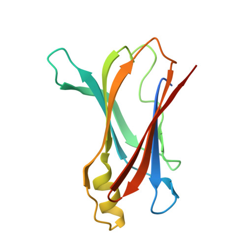
Human transthyretin in complex with iododiflunisal: structural features associated with a potent amyloid inhibitor.

Gales, L.Macedo-Ribeiro, S.Arsequell, G.Valencia, G.Saraiva, M.J.Damas, A.M.

(2005) Biochem J 388: 615-621

  • DOI: https://doi.org/10.1042/BJ20042035
  • Primary Citation Related Structures: 
    1Y1D

  • PubMed Abstract: 

    Ex vivo and in vitro studies have revealed the remarkable amyloid inhibitory potency and specificity of iododiflunisal in relation to transthyretin [Almeida, Macedo, Cardoso, Alves, Valencia, Arsequell, Planas and Saraiva (2004) Biochem. J. 381, 351-356], a protein implicated in familial amyloidotic polyneuropathy. In the present paper, the crystal structure of transthyretin complexed with this diflunisal derivative is reported, which enables a detailed analysis of the protein-ligand interactions. Iododiflunisal binds very deep in the hormone-binding channel. The iodine substituent is tightly anchored into a pocket of the binding site and the fluorine atoms provide extra hydrophobic contacts with the protein. The carboxylate substituent is involved in an electrostatic interaction with the N(zeta) of a lysine residue. Moreover, ligand-induced conformational alterations in the side chain of some residues result in the formation of new intersubunit hydrogen bonds. All these new interactions, induced by iododiflunisal, increase the stability of the tetramer impairing the formation of amyloid fibrils. The crystal structure of this complex opens perspectives for the design of more specific and effective drugs for familial amyloidotic polyneuropathy patients.


  • Organizational Affiliation
    • Instituto de Ciências Biomédicas Abel Salazar and Instituto de Biologia Molecular e Celular, Rua do Campo Alegre 823, 4150 Porto, Portugal.

Macromolecule Content 

  • Total Structure Weight: 28.31 kDa 
  • Atom Count: 1,967 
  • Modeled Residue Count: 231 
  • Deposited Residue Count: 254 
  • Unique protein chains: 1

Macromolecules

Find similar proteins by:|  3D Structure
Entity ID: 1
MoleculeChains  Sequence LengthOrganismDetailsImage
Transthyretin
A, B
127Homo sapiensMutation(s): 0 
Gene Names: TTRPALB
UniProt & NIH Common Fund Data Resources
Find proteins for P02766 (Homo sapiens)
Explore P02766 
Go to UniProtKB:  P02766
PHAROS:  P02766
GTEx:  ENSG00000118271 
Entity Groups
Sequence Clusters30% Identity50% Identity70% Identity90% Identity95% Identity100% Identity
UniProt GroupP02766
Sequence Annotations
Expand
Reference Sequence

Small Molecules

Ligands 1 Unique
IDChains Name / Formula / InChI Key2D Diagram3D Interactions
FHI

Query on FHI


Download:Ideal Coordinates CCD File
C [auth A],
D [auth B]
2',4'-DIFLUORO-4-HYDROXY-5-IODO-1,1'-BIPHENYL-3-CARBOXYLIC ACID
C13 H7 F2 I O3
SSYOLLIRAAWGDJ-UHFFFAOYSA-N

Experimental Data & Validation

Experimental Data

  • Method: X-RAY DIFFRACTION
  • Resolution: 1.70 Å
  • R-Value Free:  0.222 (Depositor), 0.224 (DCC) 
  • R-Value Work:  0.197 (Depositor), 0.202 (DCC) 
  • R-Value Observed: 0.198 (Depositor) 
Space Group: P 21 21 2
Unit Cell:
Length ( Å )Angle ( ˚ )
a = 43.313α = 90
b = 85.844β = 90
c = 64.865γ = 90
Software Package:
Software NamePurpose
MOSFLMdata reduction
SCALAdata scaling
AMoREphasing
REFMACrefinement
CCP4data scaling

Structure Validation

View Full Validation Report


Ligand Structure Quality Assessment 


Entry History 

Deposition Data

Revision History  (Full details and data files)

  • Version 1.0: 2005-07-26
    Type: Initial release
  • Version 1.1: 2008-04-30
    Changes: Version format compliance
  • Version 1.2: 2011-07-13
    Changes: Derived calculations, Version format compliance
  • Version 1.3: 2023-08-23
    Changes: Data collection, Database references, Derived calculations, Refinement description