1XTM | pdb_00001xtm

Crystal structure of the double mutant Y88H-P104H of a SOD-like protein from Bacillus subtilis.


Experimental Data Snapshot

  • Method: X-RAY DIFFRACTION
  • Resolution: 1.60 Å
  • R-Value Free: 
    0.266 (Depositor), 0.267 (DCC) 
  • R-Value Work: 
    0.253 (Depositor), 0.253 (DCC) 
  • R-Value Observed: 
    0.254 (Depositor) 

Starting Model: experimental
View more details

wwPDB Validation 3D Report Full Report

Validation slider image for 1XTM

This is version 1.5 of the entry. See complete history

Literature

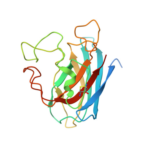
From an Inactive Prokaryotic SOD Homologue to an Active Protein through Site-Directed Mutagenesis.

Banci, L.Benvenuti, M.Bertini, I.Cabelli, D.E.Calderone, V.Fantoni, A.Mangani, S.Migliardi, M.Viezzoli, M.S.

(2005) J Am Chem Soc 127: 13287-13292

  • DOI: https://doi.org/10.1021/ja052790o
  • Primary Citation Related Structures: 
    1XTL, 1XTM

  • PubMed Abstract: 

    It is known that several prokaryotic protein sequences, characterized by high homology with the eukaryotic Cu,ZnSODs, lack some of the metal ligands. In the present work, we have stepwise reintroduced the two missing copper ligands in the SOD-like protein of Bacillus subtilis, through site-directed mutagenesis. The mutant with three out of the four His that bind copper is not active, whereas the fully reconstituted mutant displays an activity of about 10% that of human Cu,ZnSOD. The mutated proteins have been characterized in solution and in the solid state. In solution, the proteins experience conformational disorder, which is believed to be partly responsible for the decreased enzymatic activity and sheds light on the tendency of several human SOD mutants to introduce mobility in the protein frame. In the crystal, on the contrary, the protein has a well-defined conformation, giving rise to dimers through the coordination of an exogenous zinc ion. The catalytic properties of the double mutant, which might be regarded as a step in an artificial evolution from a nonactive SOD to a fully functioning enzyme, are discussed on the basis of the structural and dynamical properties.


  • Organizational Affiliation
    • Department of Chemistry and Centro Risonanze Magnetiche, University of Florence, 50019 Sesto Fiorentino, Italy.

Macromolecule Content 

  • Total Structure Weight: 37.96 kDa 
  • Atom Count: 2,507 
  • Modeled Residue Count: 305 
  • Deposited Residue Count: 350 
  • Unique protein chains: 1

Macromolecules

Find similar proteins by:|  3D Structure
Entity ID: 1
MoleculeChains  Sequence LengthOrganismDetailsImage
Hypothetical superoxide dismutase-like protein yojMA [auth B],
B [auth A]
175Bacillus subtilisMutation(s): 2 
UniProt
Find proteins for O31851 (Bacillus subtilis (strain 168))
Explore O31851 
Go to UniProtKB:  O31851
Entity Groups
Sequence Clusters30% Identity50% Identity70% Identity90% Identity95% Identity100% Identity
UniProt GroupO31851
Sequence Annotations
Expand
Reference Sequence

Experimental Data & Validation

Experimental Data

  • Method: X-RAY DIFFRACTION
  • Resolution: 1.60 Å
  • R-Value Free:  0.266 (Depositor), 0.267 (DCC) 
  • R-Value Work:  0.253 (Depositor), 0.253 (DCC) 
  • R-Value Observed: 0.254 (Depositor) 
Space Group: P 21 21 2
Unit Cell:
Length ( Å )Angle ( ˚ )
a = 52.462α = 90
b = 104.35β = 90
c = 58.756γ = 90
Software Package:
Software NamePurpose
REFMACrefinement
MOSFLMdata reduction
CCP4data scaling
MOLREPphasing

Structure Validation

View Full Validation Report


Entry History 

Deposition Data

Revision History  (Full details and data files)

  • Version 1.0: 2005-10-04
    Type: Initial release
  • Version 1.1: 2008-04-30
    Changes: Version format compliance
  • Version 1.2: 2011-07-13
    Changes: Derived calculations, Version format compliance
  • Version 1.3: 2021-11-10
    Changes: Data collection, Database references, Derived calculations
  • Version 1.4: 2023-10-25
    Changes: Data collection, Refinement description
  • Version 1.5: 2024-11-20
    Changes: Structure summary