1XSL | pdb_00001xsl

Crystal Structure of human DNA polymerase lambda in complex with a one nucleotide DNA gap


Experimental Data Snapshot

  • Method: X-RAY DIFFRACTION
  • Resolution: 2.30 Å
  • R-Value Free: 
    0.252 (Depositor), 0.244 (DCC) 
  • R-Value Work: 
    0.207 (Depositor), 0.198 (DCC) 
  • R-Value Observed: 
    0.207 (Depositor) 

wwPDB Validation 3D Report Full Report

Validation slider image for 1XSL

This is version 1.4 of the entry. See complete history

Literature

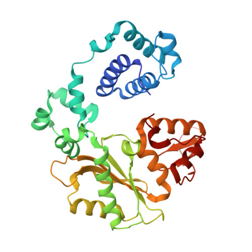
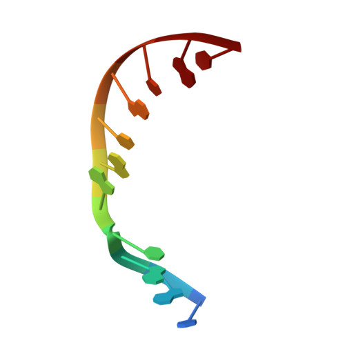
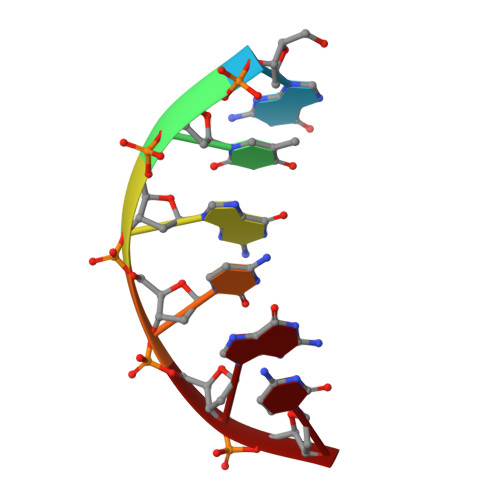
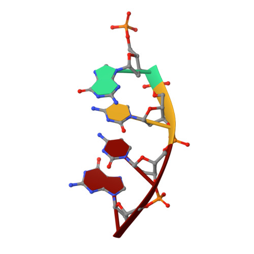
A closed conformation for the Pol lambda catalytic cycle.

Garcia-Diaz, M.Bebenek, K.Krahn, J.M.Kunkel, T.A.Pedersen, L.C.

(2005) Nat Struct Mol Biol 12: 97-98

  • DOI: https://doi.org/10.1038/nsmb876
  • Primary Citation Related Structures: 
    1XSL, 1XSN, 1XSP

  • PubMed Abstract: 

    Pol lambda is a family X member believed to fill short gaps during DNA repair. Here we report crystal structures of Pol lambda representing three steps in filling a single-nucleotide gap. These structures indicate that, unlike other DNA polymerases, Pol lambda does not undergo large subdomain movements during catalysis, and they provide a clear characterization of the geometry and stereochemistry of the in-line nucleotidyl transfer reaction.


  • Organizational Affiliation
    • Laboratory of Structural Biology, National Institute of Environmental Health Sciences, National Institutes of Health, Department of Health and Human Services, Research Triangle Park, North Carolina 27709, USA.

Macromolecule Content 

  • Total Structure Weight: 175.71 kDa 
  • Atom Count: 12,778 
  • Modeled Residue Count: 1,389 
  • Deposited Residue Count: 1,424 
  • Unique protein chains: 1
  • Unique nucleic acid chains: 3

Macromolecules


Find similar proteins by:|  3D Structure
Entity ID: 4
MoleculeChains  Sequence LengthOrganismDetailsImage
DNA polymerase lambdaM [auth A],
N [auth E],
O [auth I],
P [auth M]
335Homo sapiensMutation(s): 0 
Gene Names: POLL
EC: 2.7.7.7 (PDB Primary Data), 4.2.99 (UniProt)
UniProt & NIH Common Fund Data Resources
Find proteins for Q9UGP5 (Homo sapiens)
Explore Q9UGP5 
Go to UniProtKB:  Q9UGP5
PHAROS:  Q9UGP5
GTEx:  ENSG00000166169 
Entity Groups
Sequence Clusters30% Identity50% Identity70% Identity90% Identity95% Identity100% Identity
UniProt GroupQ9UGP5
Sequence Annotations
Expand
Reference Sequence
Find similar nucleic acids by:  Sequence
Entity ID: 1
MoleculeChains LengthOrganismImage
5'-D(*CP*GP*GP*CP*AP*GP*CP*GP*CP*AP*C)-3'A [auth B],
D [auth F],
G [auth J],
J [auth N]
11N/A
Sequence Annotations
Expand
Reference Sequence
Find similar nucleic acids by:  Sequence
Entity ID: 2
MoleculeChains LengthOrganismImage
5'-D(*GP*TP*GP*CP*GP*C)-3'B [auth C],
E [auth G],
H [auth K],
K [auth O]
6N/A
Sequence Annotations
Expand
Reference Sequence
Find similar nucleic acids by:  Sequence
Entity ID: 3
MoleculeChains LengthOrganismImage
5'-D(P*GP*CP*CP*G)-3'C [auth D],
F [auth H],
I [auth L],
L [auth P]
4N/A
Sequence Annotations
Expand
Reference Sequence

Small Molecules

Ligands 3 Unique
IDChains Name / Formula / InChI Key2D Diagram3D Interactions
CAC

Query on CAC


Download:Ideal Coordinates CCD File
R [auth D]CACODYLATE ION
C2 H6 As O2
OGGXGZAMXPVRFZ-UHFFFAOYSA-M
MG

Query on MG


Download:Ideal Coordinates CCD File
Q [auth B],
S [auth F],
T [auth H]
MAGNESIUM ION
Mg
JLVVSXFLKOJNIY-UHFFFAOYSA-N
NA

Query on NA


Download:Ideal Coordinates CCD File
U [auth J]
V [auth N]
W [auth A]
X [auth E]
Y [auth I]
U [auth J],
V [auth N],
W [auth A],
X [auth E],
Y [auth I],
Z [auth M]
SODIUM ION
Na
FKNQFGJONOIPTF-UHFFFAOYSA-N

Experimental Data & Validation

Experimental Data

  • Method: X-RAY DIFFRACTION
  • Resolution: 2.30 Å
  • R-Value Free:  0.252 (Depositor), 0.244 (DCC) 
  • R-Value Work:  0.207 (Depositor), 0.198 (DCC) 
  • R-Value Observed: 0.207 (Depositor) 
Space Group: P 21 21 2
Unit Cell:
Length ( Å )Angle ( ˚ )
a = 191.504α = 90
b = 98.812β = 90
c = 104.519γ = 90
Software Package:
Software NamePurpose
CNSrefinement
HKL-2000data reduction
SCALEPACKdata scaling
MOLREPphasing

Structure Validation

View Full Validation Report


Entry History 

Deposition Data

Revision History  (Full details and data files)

  • Version 1.0: 2005-01-18
    Type: Initial release
  • Version 1.1: 2008-04-30
    Changes: Version format compliance
  • Version 1.2: 2011-07-13
    Changes: Non-polymer description, Version format compliance
  • Version 1.3: 2018-01-24
    Changes: Database references
  • Version 1.4: 2024-11-20
    Changes: Data collection, Database references, Derived calculations, Structure summary