1XQM | pdb_00001xqm

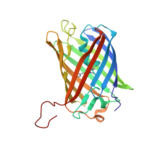
Variations on the GFP chromophore scaffold: A fragmented 5-membered heterocycle revealed in the 2.1A crystal structure of a non-fluorescent chromoprotein


Experimental Data Snapshot

  • Method: X-RAY DIFFRACTION
  • Resolution: 2.10 Å
  • R-Value Free: 
    0.233 (Depositor) 
  • R-Value Work: 
    0.214 (Depositor) 
  • R-Value Observed: 
    0.214 (Depositor) 

wwPDB Validation 3D Report Full Report

Validation slider image for 1XQM

This is version 2.0 of the entry. See complete history

Literature

Variations on the GFP chromophore: A polypeptide fragmentation within the chromophore revealed in the 2.1-A crystal structure of a nonfluorescent chromoprotein from Anemonia sulcata

Wilmann, P.G.Petersen, J.Devenish, R.J.Prescott, M.Rossjohn, J.

(2005) J Biological Chem 280: 2401-2404

  • DOI: https://doi.org/10.1074/jbc.C400484200
  • Primary Citation Related Structures: 
    1XQM

  • PubMed Abstract: 

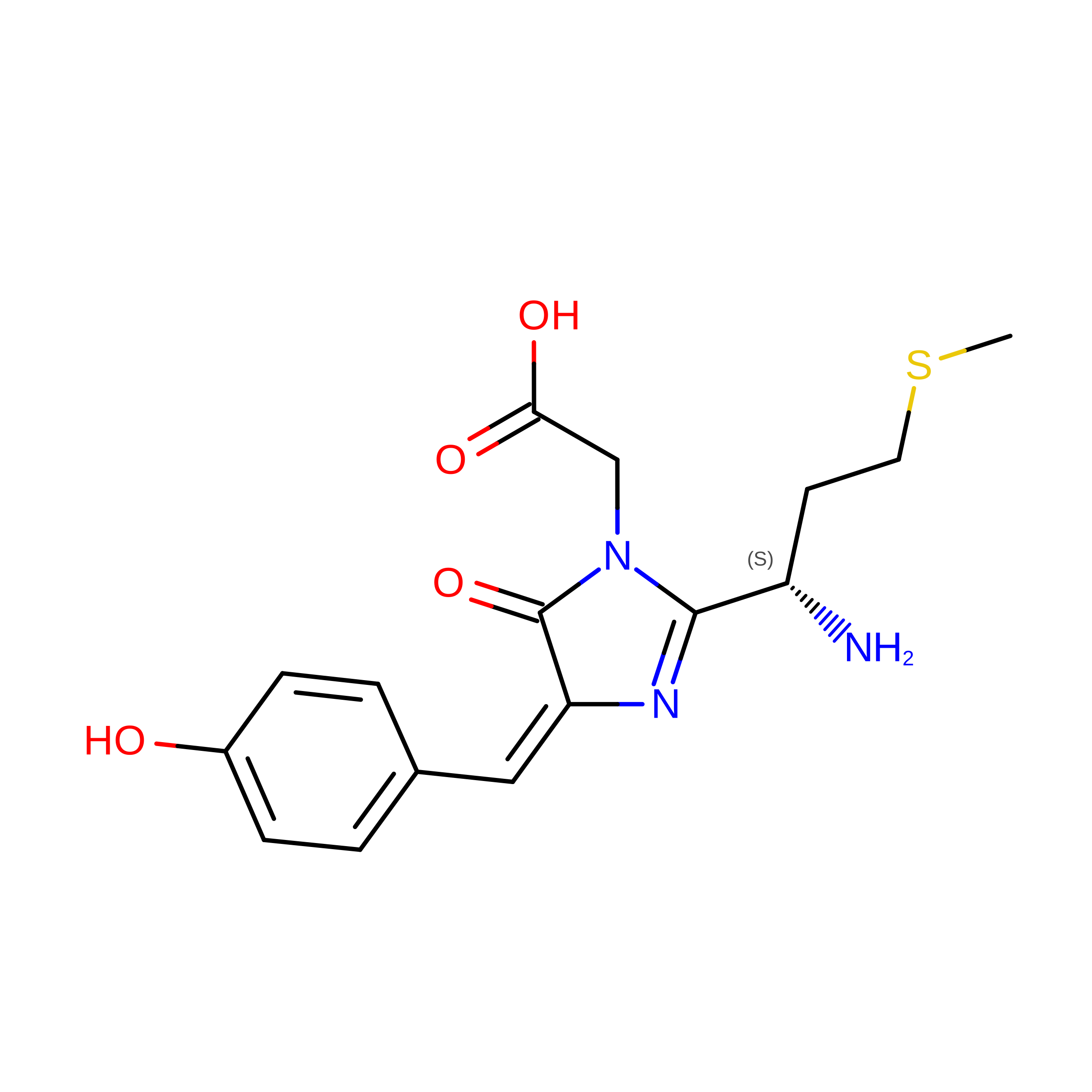
    We have determined to 2.1 A resolution the crystal structure of a dark state, kindling fluorescent protein isolated from the sea anemone, Anemonia sulcata. The chromophore sequence Met(63)-Tyr(64)-Gly(65) of the A. sulcata chromoprotein was previously proposed to comprise a 6-membered pyrazine-type heterocycle (Martynov, V. I., Savitsky, A. P., Martynova, N. Y., Savitsky, P. A., Lukyanov, K. A., and Lukyanov, S. A. (2001) J. Biol. Chem. 276, 21012-21016). However, our crystallographic data revealed the chromophore to comprise a 5-membered p-hydroxybenzylideneimidazolinone moiety that adopts a non-coplanar trans conformation within the interior of the GFP beta-can fold. Unexpectedly, fragmentation of the polypeptide was found to occur within the chromophore moiety, at the bond between Cys(62C) and Met(63N1.) Our structural data reveal that fragmentation of the chromophore represents an intrinsic, autocatalytic step toward the formation of the mature chromophore within the specific GFP-like proteins.


  • Organizational Affiliation
    • Protein Crystallography Unit, Monash Centre for Synchrotron Science.

Macromolecule Content 

  • Total Structure Weight: 25.9 kDa 
  • Atom Count: 1,924 
  • Modeled Residue Count: 228 
  • Deposited Residue Count: 230 
  • Unique protein chains: 1

Macromolecules

Find similar proteins by:|  3D Structure
Entity ID: 1
MoleculeChains  Sequence LengthOrganismDetailsImage
kindling fluorescent protein230N/AMutation(s): 1 
UniProt
Find proteins for Q9GZ28 (Anemonia sulcata)
Explore Q9GZ28 
Go to UniProtKB:  Q9GZ28
Entity Groups
Sequence Clusters30% Identity50% Identity70% Identity90% Identity95% Identity100% Identity
UniProt GroupQ9GZ28
Sequence Annotations
Expand
Reference Sequence

Small Molecules

Ligands 1 Unique
IDChains Name / Formula / InChI Key2D Diagram3D Interactions
ACY

Query on ACY


Download:Ideal Coordinates CCD File
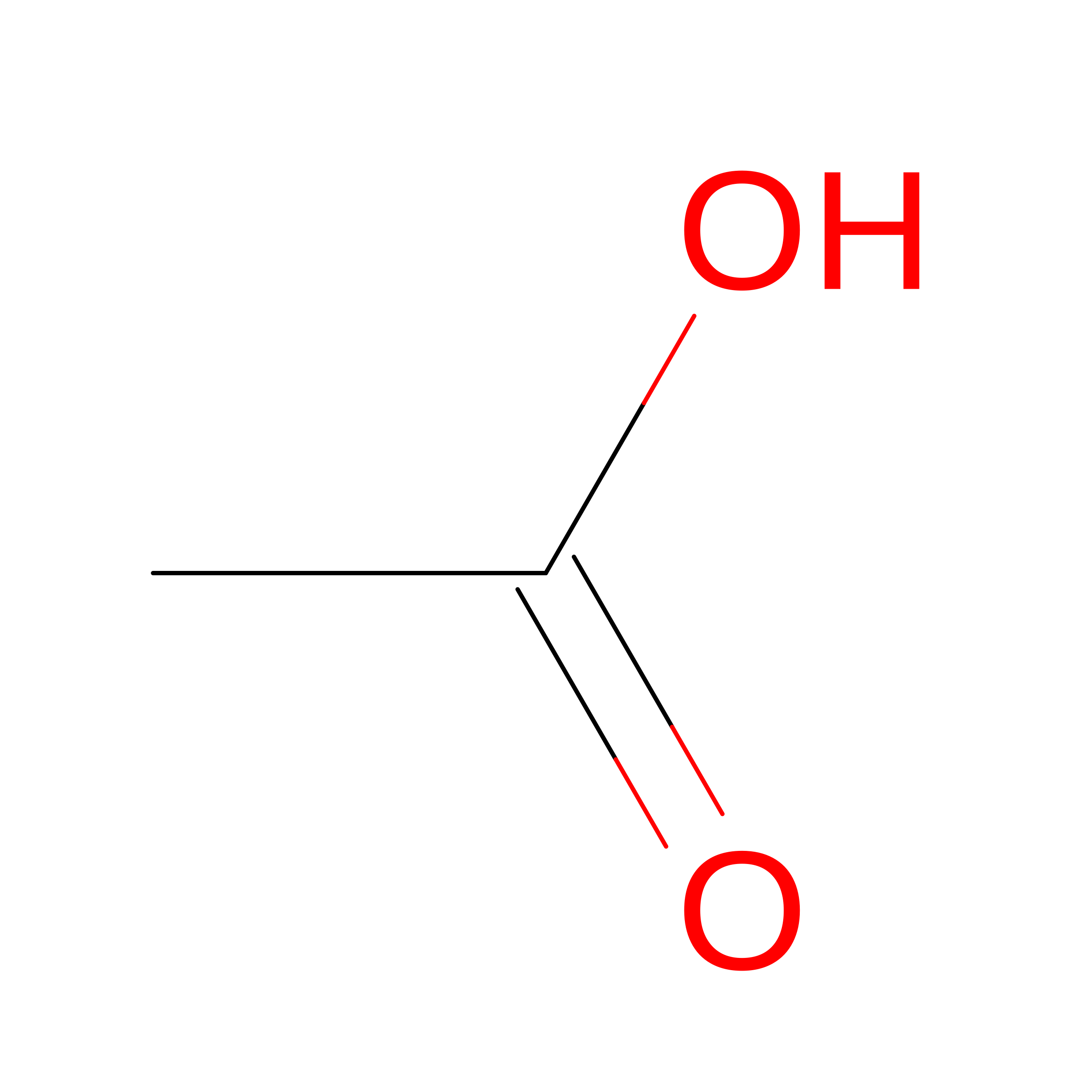
B [auth A]ACETIC ACID
C2 H4 O2
QTBSBXVTEAMEQO-UHFFFAOYSA-N
Modified Residues  1 Unique
IDChains TypeFormula2D DiagramParent
CH6
Query on CH6
A
L-PEPTIDE LINKINGC16 H19 N3 O4 SMET, TYR, GLY

Experimental Data & Validation

Experimental Data

  • Method: X-RAY DIFFRACTION
  • Resolution: 2.10 Å
  • R-Value Free:  0.233 (Depositor) 
  • R-Value Work:  0.214 (Depositor) 
  • R-Value Observed: 0.214 (Depositor) 
Space Group: P 62 2 2
Unit Cell:
Length ( Å )Angle ( ˚ )
a = 112.482α = 90
b = 112.482β = 90
c = 96.906γ = 120
Software Package:
Software NamePurpose
CNSrefinement

Structure Validation

View Full Validation Report


Entry History 

Deposition Data

Revision History  (Full details and data files)

  • Version 1.0: 2004-11-16
    Type: Initial release
  • Version 1.1: 2008-04-30
    Changes: Version format compliance
  • Version 1.2: 2011-07-13
    Changes: Derived calculations, Version format compliance
  • Version 1.3: 2024-10-09
    Changes: Advisory, Data collection, Database references, Derived calculations, Structure summary
  • Version 2.0: 2026-03-18
    Changes: Polymer sequence