1XPM | pdb_00001xpm

Crystal Structure of Staphylococcus aureus HMG-COA Synthase with HMG-CoA and Acetoacetyl-COA and Acetylated Cysteine


Experimental Data Snapshot

  • Method: X-RAY DIFFRACTION
  • Resolution: 1.60 Å
  • R-Value Free: 
    0.226 (Depositor), 0.243 (DCC) 
  • R-Value Work: 
    0.210 (Depositor), 0.229 (DCC) 

Starting Model: experimental
View more details

wwPDB Validation 3D Report Full Report

Validation slider image for 1XPM

Ligand Structure Quality Assessment 


This is version 1.5 of the entry. See complete history

Literature

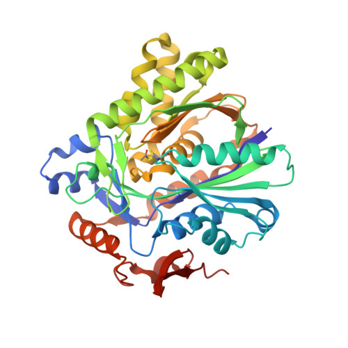
3-hydroxy-3-methylglutaryl-CoA synthase intermediate complex observed in "real-time"

Theisen, M.J.Misra, I.Saadat, D.Campobasso, N.Miziorko, H.M.Harrison, D.H.T.

(2004) Proc Natl Acad Sci U S A 47: 16442-16447

  • DOI: https://doi.org/10.1073/pnas.0405809101
  • Primary Citation Related Structures: 
    1XPK, 1XPL, 1XPM

  • PubMed Abstract: 

    The formation of carbon-carbon bonds via an acyl-enzyme intermediate plays a central role in fatty acid, polyketide, and isoprenoid biosynthesis. Uniquely among condensing enzymes, 3-hydroxy-3-methylglutaryl (HMG)-CoA synthase (HMGS) catalyzes the formation of a carbon-carbon bond by activating the methyl group of an acetylated cysteine. This reaction is essential in Gram-positive bacteria, and represents the first committed step in human cholesterol biosynthesis. Reaction kinetics, isotope exchange, and mass spectroscopy suggest surprisingly that HMGS is able to catalyze the "backwards" reaction in solution, where HMG-CoA is cleaved to form acetoacetyl-CoA (AcAc-CoA) and acetate. Here, we trap a complex of acetylated HMGS from Staphylococcus aureus and bound acetoacetyl-CoA by cryo-cooling enzyme crystals at three different times during the course of its back-reaction with its physiological product (HMG-CoA). This nonphysiological "backwards" reaction is used to understand the details of the physiological reaction with regards to individual residues involved in catalysis and substrate/product binding. The structures suggest that an active-site glutamic acid (Glu-79) acts as a general base both in the condensation between acetoacetyl-CoA and the acetylated enzyme, and the hydrolytic release of HMG-CoA from the enzyme. The ability to trap this enzyme-intermediate complex may suggest a role for protein dynamics and the interplay between protomers during the normal course of catalysis.


  • Organizational Affiliation
    • Department of Biochemistry and Molecular Biology, Rosalind Franklin University of Medicine and Science, North Chicago, IL 60064, USA.

Macromolecule Content 

  • Total Structure Weight: 185.14 kDa 
  • Atom Count: 15,324 
  • Modeled Residue Count: 1,560 
  • Deposited Residue Count: 1,588 
  • Unique protein chains: 1

Macromolecules

Find similar proteins by:|  3D Structure
Entity ID: 1
MoleculeChains  Sequence LengthOrganismDetailsImage
3-hydroxy-3-methylglutaryl CoA synthase
A, B, C, D
396Staphylococcus aureusMutation(s): 1 
Gene Names: mvaS
EC: 2.3.3.10

Small Molecules

Ligands 3 Unique
IDChains Name / Formula / InChI Key2D Diagram3D Interactions
HMG

Query on HMG


Download:Ideal Coordinates CCD File
G [auth A],
K [auth B],
O [auth C],
S [auth D]
3-HYDROXY-3-METHYLGLUTARYL-COENZYME A
C27 H39 N7 O20 P3 S
CABVTRNMFUVUDM-VRHQGPGLSA-I
CAA

Query on CAA


Download:Ideal Coordinates CCD File
H [auth A],
L [auth B],
P [auth C],
T [auth D]
ACETOACETYL-COENZYME A
C25 H40 N7 O18 P3 S
OJFDKHTZOUZBOS-CITAKDKDSA-N
SO4

Query on SO4


Download:Ideal Coordinates CCD File
E [auth A]
F [auth A]
I [auth B]
J [auth B]
M [auth C]
E [auth A],
F [auth A],
I [auth B],
J [auth B],
M [auth C],
N [auth C],
Q [auth D],
R [auth D]
SULFATE ION
O4 S
QAOWNCQODCNURD-UHFFFAOYSA-L
Modified Residues  1 Unique
IDChains TypeFormula2D DiagramParent
SCY
Query on SCY
A, B, C, D
L-PEPTIDE LINKINGC5 H9 N O3 SCYS

Experimental Data & Validation

Experimental Data

  • Method: X-RAY DIFFRACTION
  • Resolution: 1.60 Å
  • R-Value Free:  0.226 (Depositor), 0.243 (DCC) 
  • R-Value Work:  0.210 (Depositor), 0.229 (DCC) 
Space Group: P 1 21 1
Unit Cell:
Length ( Å )Angle ( ˚ )
a = 65.119α = 90
b = 118.774β = 90.08
c = 121.304γ = 90
Software Package:
Software NamePurpose
DENZOdata reduction
SCALEPACKdata scaling
CNSrefinement
PDB_EXTRACTdata extraction
HKL-2000data reduction
BEASTphasing

Structure Validation

View Full Validation Report


Ligand Structure Quality Assessment 


Entry History 

Deposition Data

Revision History  (Full details and data files)

  • Version 1.0: 2004-11-02
    Type: Initial release
  • Version 1.1: 2008-04-30
    Changes: Version format compliance
  • Version 1.2: 2011-07-13
    Changes: Version format compliance
  • Version 1.3: 2017-10-11
    Changes: Refinement description
  • Version 1.4: 2023-08-23
    Changes: Data collection, Database references, Derived calculations, Refinement description
  • Version 1.5: 2024-10-30
    Changes: Structure summary