1XP5 | pdb_00001xp5

Structure Of The (Sr)Ca2+-ATPase E2-AlF4- Form


Experimental Data Snapshot

  • Method: X-RAY DIFFRACTION
  • Resolution: 3.00 Å
  • R-Value Free: 
    0.265 (Depositor), 0.236 (DCC) 
  • R-Value Work: 
    0.237 (Depositor), 0.235 (DCC) 
  • R-Value Observed: 
    0.245 (Depositor) 

Starting Model: other
View more details

wwPDB Validation 3D Report Full Report

Validation slider image for 1XP5

Ligand Structure Quality Assessment 


This is version 1.4 of the entry. See complete history

Literature

Dephosphorylation of the Calcium Pump Coupled to Counterion Occlusion

Olesen, C.Sorensen, T.L.S.Nielsen, R.C.Moller, J.V.Nissen, P.

(2004) Science 306: 2251-2255

  • DOI: https://doi.org/10.1126/science.1106289
  • Primary Citation Related Structures: 
    1XP5

  • PubMed Abstract: 

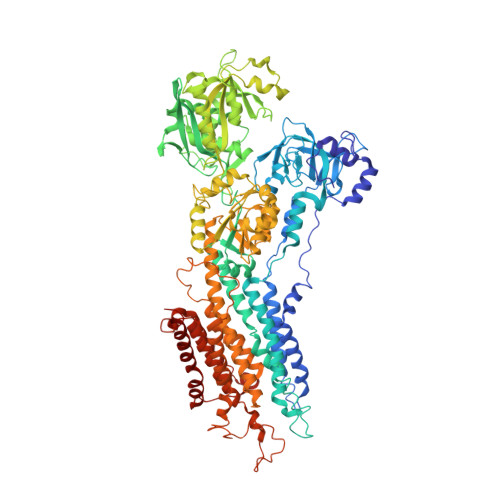
    P-type ATPases extract energy by hydrolysis of adenosine triphosphate (ATP) in two steps, formation and breakdown of a covalent phosphoenzyme intermediate. This process drives active transport and countertransport of the cation pumps. We have determined the crystal structure of rabbit sarcoplasmic reticulum Ca2+ adenosine triphosphatase in complex with aluminum fluoride, which mimics the transition state of hydrolysis of the counterion-bound (protonated) phosphoenzyme. On the basis of structural analysis and biochemical data, we find this form to represent an occluded state of the proton counterions. Hydrolysis is catalyzed by the conserved Thr-Gly-Glu-Ser motif, and it exploits an associative nucleophilic reaction mechanism of the same type as phosphoryl transfer from ATP. On this basis, we propose a general mechanism of occluded transition states of Ca2+ transport and H+ countertransport coupled to phosphorylation and dephosphorylation, respectively.


  • Organizational Affiliation
    • Centre for Structural Biology, Department of Molecular Biology, University of Aarhus, Gustav Wieds Vej 10C, DK-8000 Aarhus C, Denmark.

Macromolecule Content 

  • Total Structure Weight: 110.42 kDa 
  • Atom Count: 7,727 
  • Modeled Residue Count: 994 
  • Deposited Residue Count: 994 
  • Unique protein chains: 1

Macromolecules

Find similar proteins by:|  3D Structure
Entity ID: 1
MoleculeChains  Sequence LengthOrganismDetailsImage
Sarcoplasmic/endoplasmic reticulum calcium ATPase 1994Oryctolagus cuniculusMutation(s): 0 
EC: 3.6.3.8 (PDB Primary Data), 7.2.2.10 (UniProt)
Membrane Entity: Yes 
UniProt
Find proteins for P04191 (Oryctolagus cuniculus)
Explore P04191 
Go to UniProtKB:  P04191
Entity Groups
Sequence Clusters30% Identity50% Identity70% Identity90% Identity95% Identity100% Identity
UniProt GroupP04191
Sequence Annotations
Expand
Reference Sequence

Small Molecules

Ligands 4 Unique
IDChains Name / Formula / InChI Key2D Diagram3D Interactions
TG1

Query on TG1


Download:Ideal Coordinates CCD File
E [auth A]OCTANOIC ACID [3S-[3ALPHA, 3ABETA, 4ALPHA, 6BETA, 6ABETA, 7BETA, 8ALPHA(Z), 9BALPHA]]-6-(ACETYLOXY)-2,3,-3A,4,5,6,6A,7,8,9B-DECAHYDRO-3,3A-DIHYDROXY-3,6,9-TRIMETHYL-8-[(2-METHYL-1-OXO-2-BUTENYL)OX Y]-2-OXO-4-(1-OXOBUTOXY)-AZULENO[4,5-B]FURAN-7-YL ESTER
C34 H50 O12
IXFPJGBNCFXKPI-FSIHEZPISA-N
ALF

Query on ALF


Download:Ideal Coordinates CCD File
C [auth A]TETRAFLUOROALUMINATE ION
Al F4
UYOMQIYKOOHAMK-UHFFFAOYSA-J
K

Query on K


Download:Ideal Coordinates CCD File
D [auth A]POTASSIUM ION
K
NPYPAHLBTDXSSS-UHFFFAOYSA-N
MG

Query on MG


Download:Ideal Coordinates CCD File
B [auth A]MAGNESIUM ION
Mg
JLVVSXFLKOJNIY-UHFFFAOYSA-N

Experimental Data & Validation

Experimental Data

  • Method: X-RAY DIFFRACTION
  • Resolution: 3.00 Å
  • R-Value Free:  0.265 (Depositor), 0.236 (DCC) 
  • R-Value Work:  0.237 (Depositor), 0.235 (DCC) 
  • R-Value Observed: 0.245 (Depositor) 
Space Group: P 21 21 2
Unit Cell:
Length ( Å )Angle ( ˚ )
a = 86.51α = 90
b = 119.27β = 90
c = 142.26γ = 90
Software Package:
Software NamePurpose
DENZOdata reduction
SCALEPACKdata scaling
PHASERphasing
CNSrefinement

Structure Validation

View Full Validation Report


Ligand Structure Quality Assessment 


Entry History 

Deposition Data

Revision History  (Full details and data files)

  • Version 1.0: 2005-01-04
    Type: Initial release
  • Version 1.1: 2008-04-30
    Changes: Version format compliance
  • Version 1.2: 2011-07-13
    Changes: Non-polymer description, Version format compliance
  • Version 1.3: 2024-03-13
    Changes: Data collection, Database references, Derived calculations, Structure summary
  • Version 1.4: 2024-04-03
    Changes: Refinement description