1XKZ | pdb_00001xkz

Crystal structure of the acylated beta-lactam sensor domain of Blar1 from S. aureus


Experimental Data Snapshot

  • Method: X-RAY DIFFRACTION
  • Resolution: 1.75 Å
  • R-Value Free: 
    0.234 (Depositor), 0.234 (DCC) 
  • R-Value Work: 
    0.205 (Depositor), 0.205 (DCC) 
  • R-Value Observed: 
    0.209 (Depositor) 

Starting Model: experimental
View more details

wwPDB Validation 3D Report Full Report

Validation slider image for 1XKZ

Ligand Structure Quality Assessment 


This is version 1.4 of the entry. See complete history

Literature

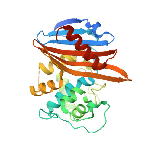
X-ray crystal structure of the acylated beta-lactam sensor domain of BlaR1 from Staphylococcus aureus and the mechanism of receptor activation for signal transduction

Birck, C.Cha, J.Y.Cross, J.Schulze-Briese, C.Meroueh, S.O.Schlegel, H.B.Mobashery, S.Samama, J.-P.

(2004) J Am Chem Soc 126: 13945-13947

  • DOI: https://doi.org/10.1021/ja044742u
  • Primary Citation Related Structures: 
    1XKZ

  • PubMed Abstract: 

    Methicillin-resistant strains of Staphylococcus aureus (MRSA) are the major cause of infections worldwide. Transcription of the beta-lactamase and PBP2a resistance genes is mediated by two closely related signal-transducing integral membrane proteins, BlaR1 and MecR1, upon binding of the beta-lactam inducer to the sensor domain. Herein we report the crystal structure at 1.75 A resolution of the sensor domain of BlaR1 in complex with a cephalosporin antibiotic. Activation of the signal transducer involves acylation of serine 389 by the beta-lactam antibiotic, a process promoted by the N-carboxylated side chain of Lys392. We present evidence that, on acylation, the lysine side chain experiences a spontaneous decarboxylation that entraps the sensor in its activated state. Kinetic determinations and quantum mechanical/molecular mechanical calculations and the interaction networks in the crystal structure shed light on how this unprecedented process for activation of a receptor may be achieved and provide insights into the mechanistic features that differentiate the signal-transducing receptor from the structurally related class D beta-lactamases, enzymes of antibiotic resistance.


  • Organizational Affiliation
    • Département de Génomique et Biologie Structurales, IGBMC CNRS/INSERM/ULP, 1 rue Laurent Fries, BP 10142, 67404 - Illkirch CU Strasbourg, France.

Macromolecule Content 

  • Total Structure Weight: 122.32 kDa 
  • Atom Count: 8,056 
  • Modeled Residue Count: 983 
  • Deposited Residue Count: 1,020 
  • Unique protein chains: 1

Macromolecules

Find similar proteins by:|  3D Structure
Entity ID: 1
MoleculeChains  Sequence LengthOrganismDetailsImage
Regulatory protein blaR1
A, B, C, D
255Staphylococcus aureusMutation(s): 0 
UniProt
Find proteins for P18357 (Staphylococcus aureus)
Explore P18357 
Go to UniProtKB:  P18357
Entity Groups
Sequence Clusters30% Identity50% Identity70% Identity90% Identity95% Identity100% Identity
UniProt GroupP18357
Sequence Annotations
Expand
Reference Sequence

Small Molecules

Ligands 3 Unique
IDChains Name / Formula / InChI Key2D Diagram3D Interactions
CAZ

Query on CAZ


Download:Ideal Coordinates CCD File
E [auth A],
H [auth B],
J [auth C],
K [auth D]
ACYLATED CEFTAZIDIME
C17 H19 N5 O7 S2
VEHPZKIFULQYFS-BZXVCXBKSA-N
EPE

Query on EPE


Download:Ideal Coordinates CCD File
I [auth B]4-(2-HYDROXYETHYL)-1-PIPERAZINE ETHANESULFONIC ACID
C8 H18 N2 O4 S
JKMHFZQWWAIEOD-UHFFFAOYSA-N
SO4

Query on SO4


Download:Ideal Coordinates CCD File
F [auth B],
G [auth B]
SULFATE ION
O4 S
QAOWNCQODCNURD-UHFFFAOYSA-L

Experimental Data & Validation

Experimental Data

  • Method: X-RAY DIFFRACTION
  • Resolution: 1.75 Å
  • R-Value Free:  0.234 (Depositor), 0.234 (DCC) 
  • R-Value Work:  0.205 (Depositor), 0.205 (DCC) 
  • R-Value Observed: 0.209 (Depositor) 
Space Group: P 1 21 1
Unit Cell:
Length ( Å )Angle ( ˚ )
a = 59.205α = 90
b = 109.791β = 106.13
c = 91.605γ = 90
Software Package:
Software NamePurpose
MOSFLMdata reduction
SCALAdata scaling
CNSrefinement
CCP4data scaling
CNSphasing

Structure Validation

View Full Validation Report


Ligand Structure Quality Assessment 


Entry History 

Deposition Data

Revision History  (Full details and data files)

  • Version 1.0: 2004-11-30
    Type: Initial release
  • Version 1.1: 2008-04-30
    Changes: Version format compliance
  • Version 1.2: 2011-07-13
    Changes: Version format compliance
  • Version 1.3: 2024-04-03
    Changes: Data collection, Database references, Derived calculations, Refinement description
  • Version 1.4: 2024-11-13
    Changes: Structure summary