1XJB | pdb_00001xjb

Crystal structure of human type 3 3alpha-hydroxysteroid dehydrogenase in complex with NADP(H), citrate and acetate molecules


Experimental Data Snapshot

  • Method: X-RAY DIFFRACTION
  • Resolution: 1.90 Å
  • R-Value Free: 
    0.199 (Depositor), 0.193 (DCC) 
  • R-Value Work: 
    0.174 (Depositor), 0.179 (DCC) 
  • R-Value Observed: 
    0.175 (Depositor) 

Starting Model: experimental
View more details

wwPDB Validation 3D Report Full Report

Validation slider image for 1XJB

Ligand Structure Quality Assessment 


This is version 1.4 of the entry. See complete history

Literature

Comparison of crystal structures of human type 3 3alpha-hydroxysteroid dehydrogenase reveals an "induced-fit" mechanism and a conserved basic motif involved in the binding of androgen

Couture, J.-F.Roy, A.-M.Cantin, L.Legrand, P.Luu-The, V.Labrie, F.Breton, R.

(2005) Protein Sci 14: 1485-1497

  • DOI: https://doi.org/10.1110/ps.051353205
  • Primary Citation Related Structures: 
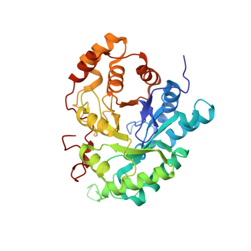
    1XJB

  • PubMed Abstract: 

    The aldo-keto reductase (AKR) human type 3 3alpha-hydroxysteroid dehydrogenase (h3alpha-HSD3, AKR1C2) plays a crucial role in the regulation of the intracellular concentrations of testosterone and 5alpha-dihydrotestosterone (5alpha-DHT), two steroids directly linked to the etiology and the progression of many prostate diseases and cancer. This enzyme also binds many structurally different molecules such as 4-hydroxynonenal, polycyclic aromatic hydrocarbons, and indanone. To understand the mechanism underlying the plasticity of its substrate-binding site, we solved the binary complex structure of h3alpha-HSD3-NADP(H) at 1.9 A resolution. During the refinement process, we found acetate and citrate molecules deeply engulfed in the steroid-binding cavity. Superimposition of this structure with the h3alpha-HSD3-NADP(H)-testosterone/acetate ternary complex structure reveals that one of the mobile loops forming the binding cavity operates a slight contraction movement against the citrate molecule while the side chains of many residues undergo numerous conformational changes, probably to create an optimal binding site for the citrate. These structural changes, which altogether cause a reduction of the substrate-binding cavity volume (from 776 A(3) in the presence of testosterone/acetate to 704 A(3) in the acetate/citrate complex), are reminiscent of the "induced-fit" mechanism previously proposed for the aldose reductase, another member of the AKR superfamily. We also found that the replacement of residues Arg(301) and Arg(304), localized near the steroid-binding cavity, significantly affects the 3alpha-HSD activity of this enzyme toward 5alpha-DHT and completely abolishes its 17beta-HSD activity on 4-dione. All these results have thus been used to reevaluate the binding mode of this enzyme for androgens.


  • Organizational Affiliation
    • Oncology and Molecular Endocrinology Research Center, Laval University Medical Center and Laval University, Québec, Canada.

Macromolecule Content 

  • Total Structure Weight: 76.16 kDa 
  • Atom Count: 5,865 
  • Modeled Residue Count: 647 
  • Deposited Residue Count: 650 
  • Unique protein chains: 1

Macromolecules

Find similar proteins by:|  3D Structure
Entity ID: 1
MoleculeChains  Sequence LengthOrganismDetailsImage
Aldo-keto reductase family 1 member C2
A, B
325Homo sapiensMutation(s): 0 
Gene Names: AKR1C2DDH2
EC: 1.1.1.213 (PDB Primary Data), 1.1.1.209 (UniProt), 1.3.1.20 (UniProt), 1.1.1.51 (UniProt), 1.1.1.62 (UniProt), 1.1.1.357 (UniProt), 1.1.1.112 (UniProt)
UniProt & NIH Common Fund Data Resources
Find proteins for P52895 (Homo sapiens)
Explore P52895 
Go to UniProtKB:  P52895
PHAROS:  P52895
GTEx:  ENSG00000151632 
Entity Groups
Sequence Clusters30% Identity50% Identity70% Identity90% Identity95% Identity100% Identity
UniProt GroupP52895
Sequence Annotations
Expand
Reference Sequence

Small Molecules

Ligands 6 Unique
IDChains Name / Formula / InChI Key2D Diagram3D Interactions
NAP

Query on NAP


Download:Ideal Coordinates CCD File
E [auth A],
L [auth B]
NADP NICOTINAMIDE-ADENINE-DINUCLEOTIDE PHOSPHATE
C21 H28 N7 O17 P3
XJLXINKUBYWONI-NNYOXOHSSA-N
CIT

Query on CIT


Download:Ideal Coordinates CCD File
G [auth A]CITRIC ACID
C6 H8 O7
KRKNYBCHXYNGOX-UHFFFAOYSA-N
SO4

Query on SO4


Download:Ideal Coordinates CCD File
J [auth B],
K [auth B]
SULFATE ION
O4 S
QAOWNCQODCNURD-UHFFFAOYSA-L
BME

Query on BME


Download:Ideal Coordinates CCD File
H [auth A]BETA-MERCAPTOETHANOL
C2 H6 O S
DGVVWUTYPXICAM-UHFFFAOYSA-N
EDO

Query on EDO


Download:Ideal Coordinates CCD File
F [auth A],
M [auth B]
1,2-ETHANEDIOL
C2 H6 O2
LYCAIKOWRPUZTN-UHFFFAOYSA-N
ACT

Query on ACT


Download:Ideal Coordinates CCD File
C [auth A],
D [auth A],
I [auth B]
ACETATE ION
C2 H3 O2
QTBSBXVTEAMEQO-UHFFFAOYSA-M

Experimental Data & Validation

Experimental Data

  • Method: X-RAY DIFFRACTION
  • Resolution: 1.90 Å
  • R-Value Free:  0.199 (Depositor), 0.193 (DCC) 
  • R-Value Work:  0.174 (Depositor), 0.179 (DCC) 
  • R-Value Observed: 0.175 (Depositor) 
Space Group: H 3 2
Unit Cell:
Length ( Å )Angle ( ˚ )
a = 143.12α = 90
b = 143.12β = 90
c = 204.31γ = 120
Software Package:
Software NamePurpose
REFMACrefinement
XDSdata reduction
AMoREphasing
CNSrefinement
Crystaldata collection
CrystalCleardata reduction
XDSdata scaling

Structure Validation

View Full Validation Report


Ligand Structure Quality Assessment 


Entry History 

Deposition Data

Revision History  (Full details and data files)

  • Version 1.0: 2005-06-21
    Type: Initial release
  • Version 1.1: 2008-04-30
    Changes: Version format compliance
  • Version 1.2: 2011-07-13
    Changes: Advisory, Version format compliance
  • Version 1.3: 2019-07-24
    Changes: Data collection, Refinement description
  • Version 1.4: 2023-08-23
    Changes: Data collection, Database references, Derived calculations, Refinement description