1XIO | pdb_00001xio

Anabaena sensory rhodopsin


Experimental Data Snapshot

  • Method: X-RAY DIFFRACTION
  • Resolution: 2.00 Å
  • R-Value Free: 
    0.256 (Depositor), 0.272 (DCC) 
  • R-Value Work: 
    0.230 (Depositor), 0.249 (DCC) 

Starting Model: experimental
View more details

wwPDB Validation 3D Report Full Report

Validation slider image for 1XIO

Ligand Structure Quality Assessment 


This is version 2.0 of the entry. See complete history

Literature

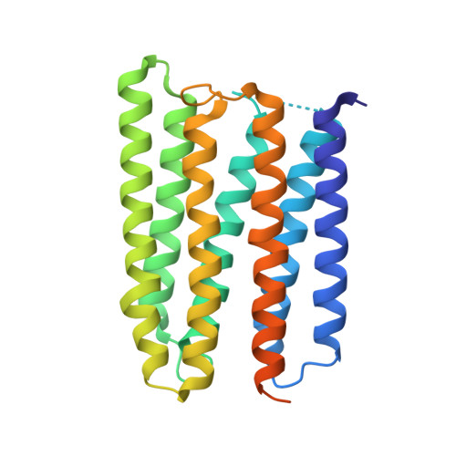
Anabaena sensory rhodopsin: a photochromic color sensor at 2.0 A.

Vogeley, L.Sineshchekov, O.A.Trivedi, V.D.Sasaki, J.Spudich, J.L.Luecke, H.

(2004) Science 306: 1390-1393

  • DOI: https://doi.org/10.1126/science.1103943
  • Primary Citation Related Structures: 
    1XIO

  • PubMed Abstract: 

    Microbial sensory rhodopsins are a family of membrane-embedded photoreceptors in prokaryotic and eukaryotic organisms. Structures of archaeal rhodopsins, which function as light-driven ion pumps or photosensors, have been reported. We present the structure of a eubacterial rhodopsin, which differs from those of previously characterized archaeal rhodopsins in its chromophore and cytoplasmic-side portions. Anabaena sensory rhodopsin exhibits light-induced interconversion between stable 13-cis and all-trans states of the retinylidene protein. The ratio of its cis and trans chromophore forms depends on the wavelength of illumination, thus providing a mechanism for a single protein to signal the color of light, for example, to regulate color-sensitive processes such as chromatic adaptation in photosynthesis. Its cytoplasmic half channel, highly hydrophobic in the archaeal rhodopsins, contains numerous hydrophilic residues networked by water molecules, providing a connection from the photoactive site to the cytoplasmic surface believed to interact with the receptor's soluble 14-kilodalton transducer.


  • Organizational Affiliation
    • Department of Molecular Biology and Biochemistry, University of California, Irvine, CA 92697, USA.

Macromolecule Content 

  • Total Structure Weight: 40.17 kDa 
  • Atom Count: 2,107 
  • Modeled Residue Count: 217 
  • Deposited Residue Count: 261 
  • Unique protein chains: 1

Macromolecules

Find similar proteins by:|  3D Structure
Entity ID: 1
MoleculeChains  Sequence LengthOrganismDetailsImage
ANABAENA SENSORY RHODOPSIN261Nostoc sp. PCC 7120 = FACHB-418Mutation(s): 0 
UniProt
Find proteins for A0ACD6B968 (Nostoc sp. (strain PCC 7120 / SAG 25.82 / UTEX 2576))
Explore A0ACD6B968 
Go to UniProtKB:  A0ACD6B968
Entity Groups
Sequence Clusters30% Identity50% Identity70% Identity90% Identity95% Identity100% Identity
UniProt GroupA0ACD6B968
Sequence Annotations
Expand
Reference Sequence

Small Molecules

Ligands 2 Unique
IDChains Name / Formula / InChI Key2D Diagram3D Interactions
PEE

Query on PEE


Download:Ideal Coordinates CCD File
C [auth A]
D [auth A]
E [auth A]
F [auth A]
G [auth A]
C [auth A],
D [auth A],
E [auth A],
F [auth A],
G [auth A],
H [auth A],
I [auth A],
J [auth A],
K [auth A],
L [auth A],
M [auth A],
N [auth A],
O [auth A]
1,2-dioleoyl-sn-glycero-3-phosphoethanolamine
C41 H78 N O8 P
MWRBNPKJOOWZPW-NYVOMTAGSA-N
RET

Query on RET


Download:Ideal Coordinates CCD File
B [auth A]RETINAL
C20 H28 O
NCYCYZXNIZJOKI-OVSJKPMPSA-N

Experimental Data & Validation

Experimental Data

  • Method: X-RAY DIFFRACTION
  • Resolution: 2.00 Å
  • R-Value Free:  0.256 (Depositor), 0.272 (DCC) 
  • R-Value Work:  0.230 (Depositor), 0.249 (DCC) 
Space Group: C 2 2 21
Unit Cell:
Length ( Å )Angle ( ˚ )
a = 103.095α = 90
b = 116.513β = 90
c = 56.186γ = 90
Software Package:
Software NamePurpose
DENZOdata reduction
SCALEPACKdata scaling
CNSrefinement
HKL-2000data reduction
CNSphasing

Structure Validation

View Full Validation Report


Ligand Structure Quality Assessment 


Entry History 

Deposition Data

Revision History  (Full details and data files)

  • Version 1.0: 2004-10-05
    Type: Initial release
  • Version 1.1: 2008-04-30
    Changes: Version format compliance
  • Version 1.2: 2011-07-13
    Changes: Derived calculations, Version format compliance
  • Version 1.3: 2014-10-29
    Changes: Non-polymer description
  • Version 1.4: 2017-10-11
    Changes: Refinement description
  • Version 2.0: 2023-08-23
    Changes: Advisory, Atomic model, Data collection, Database references, Derived calculations, Non-polymer description, Refinement description, Structure summary