1XIG | pdb_00001xig

MODES OF BINDING SUBSTRATES AND THEIR ANALOGUES TO THE ENZYME D-XYLOSE ISOMERASE


Experimental Data Snapshot

  • Method: X-RAY DIFFRACTION
  • Resolution: 1.70 Å
  • R-Value Observed: 
    0.162 (Depositor) 

wwPDB Validation 3D Report Full Report

Validation slider image for 1XIG

This is version 1.4 of the entry. See complete history

Literature

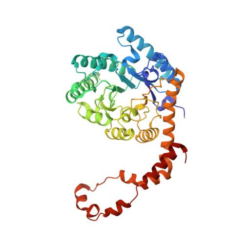
Modes of binding substrates and their analogues to the enzyme D-xylose isomerase.

Carrell, H.L.Hoier, H.Glusker, J.P.

(1994) Acta Crystallogr D Biol Crystallogr 50: 113-123

  • DOI: https://doi.org/10.1107/S0907444993009345
  • Primary Citation Related Structures: 
    1XIB, 1XIC, 1XID, 1XIE, 1XIF, 1XIG, 1XIH, 1XII, 1XIJ

  • PubMed Abstract: 

    Studies of binding of substrates and inhibitors of the enzyme D-xylose isomerase show, from X-ray diffraction data at 1.6-1.9 A resolution, that there are a variety of binding modes. These vary in the manner in which the substrate or its analogue extend, on binding, across the carboxy end of the (betaalpha)(8)-barrel structure. These binding sites are His54 and the metal ion (magnesium or manganese) that is held in place by Glul81, Asp245, Glu217 and Asp287. Possible catalytic groups have been identified in proposed mechanisms and their role in the binding of ligands is illustrated.


  • Organizational Affiliation
    • The Institute for Cancer Research, The Fox Chase Cancer Center, Philadelphia, PA 19111, USA.

Macromolecule Content 

  • Total Structure Weight: 43.52 kDa 
  • Atom Count: 3,339 
  • Modeled Residue Count: 385 
  • Deposited Residue Count: 388 
  • Unique protein chains: 1

Macromolecules

Find similar proteins by:|  3D Structure
Entity ID: 1
MoleculeChains  Sequence LengthOrganismDetailsImage
D-XYLOSE ISOMERASE388Streptomyces pseudogriseolusMutation(s): 0 
EC: 5.3.1.5
UniProt
Find proteins for P24300 (Streptomyces pseudogriseolus)
Explore P24300 
Go to UniProtKB:  P24300
Entity Groups
Sequence Clusters30% Identity50% Identity70% Identity90% Identity95% Identity100% Identity
UniProt GroupP24300
Sequence Annotations
Expand
Reference Sequence

Experimental Data & Validation

Experimental Data

  • Method: X-RAY DIFFRACTION
  • Resolution: 1.70 Å
  • R-Value Observed: 0.162 (Depositor) 
Space Group: I 2 2 2
Unit Cell:
Length ( Å )Angle ( ˚ )
a = 93.9α = 90
b = 99.7β = 90
c = 102.9γ = 90
Software Package:
Software NamePurpose
PROLSQrefinement

Structure Validation

View Full Validation Report


Entry History 

Deposition Data

Revision History  (Full details and data files)

  • Version 1.0: 1994-06-22
    Type: Initial release
  • Version 1.1: 2008-03-24
    Changes: Version format compliance
  • Version 1.2: 2011-07-13
    Changes: Derived calculations, Version format compliance
  • Version 1.3: 2020-07-29
    Type: Remediation
    Reason: Carbohydrate remediation
    Changes: Advisory, Database references, Derived calculations, Other, Structure summary
  • Version 1.4: 2024-02-14
    Changes: Data collection, Database references, Structure summary