1XEF | pdb_00001xef

Crystal structure of the ATP/Mg2+ bound composite dimer of HlyB-NBD


Experimental Data Snapshot

  • Method: X-RAY DIFFRACTION
  • Resolution: 2.50 Å
  • R-Value Free: 
    0.264 (Depositor), 0.291 (DCC) 
  • R-Value Work: 
    0.219 (Depositor) 
  • R-Value Observed: 
    0.222 (Depositor) 

Starting Model: experimental
View more details

wwPDB Validation 3D Report Full Report

Validation slider image for 1XEF

Ligand Structure Quality Assessment 


This is version 1.4 of the entry. See complete history

Literature

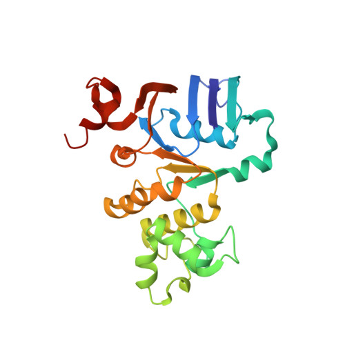
H662 is the linchpin of ATP hydrolysis in the nucleotide-binding domain of the ABC transporter HlyB

Zaitseva, J.Jenewein, S.Jumpertz, T.Holland, I.B.Schmitt, L.

(2005) EMBO J 24: 1901-1910

  • DOI: https://doi.org/10.1038/sj.emboj.7600657
  • Primary Citation Related Structures: 
    1XEF

  • PubMed Abstract: 

    The ABC transporter HlyB is a central element of the HlyA secretion machinery, a paradigm of Type I secretion. Here, we describe the crystal structure of the HlyB-NBD (nucleotide-binding domain) with H662 replaced by Ala in complex with ATP/Mg2+. The dimer shows a composite architecture, in which two intact ATP molecules are bound at the interface of the Walker A motif and the C-loop, provided by the two monomers. ATPase measurements confirm that H662 is essential for activity. Based on these data, we propose a model in which E631 and H662, highly conserved among ABC transporters, form a catalytic dyad. Here, H662 acts as a 'linchpin', holding together all required parts of a complicated network of interactions between ATP, water molecules, Mg2+, and amino acids both in cis and trans, necessary for intermonomer communication. Based on biochemical experiments, we discuss the hypothesis that substrate-assisted catalysis, rather than general base catalysis might operate in ABC-ATPases.


  • Organizational Affiliation
    • Institute of Biochemistry, Biocenter, Johann-Wolfgang Goethe University Frankfurt, Frankfurt, Germany.

Macromolecule Content 

  • Total Structure Weight: 109.55 kDa 
  • Atom Count: 7,852 
  • Modeled Residue Count: 964 
  • Deposited Residue Count: 964 
  • Unique protein chains: 1

Macromolecules

Find similar proteins by:|  3D Structure
Entity ID: 1
MoleculeChains  Sequence LengthOrganismDetailsImage
Alpha-hemolysin translocation ATP-binding protein hlyB
A, B, C, D
241Escherichia coliMutation(s): 1 
UniProt
Find proteins for P08716 (Escherichia coli)
Explore P08716 
Go to UniProtKB:  P08716
Entity Groups
Sequence Clusters30% Identity50% Identity70% Identity90% Identity95% Identity100% Identity
UniProt GroupP08716
Sequence Annotations
Expand
Reference Sequence

Small Molecules

Ligands 2 Unique
IDChains Name / Formula / InChI Key2D Diagram3D Interactions
ATP

Query on ATP


Download:Ideal Coordinates CCD File
F [auth A],
H [auth B],
J [auth C],
L [auth D]
ADENOSINE-5'-TRIPHOSPHATE
C10 H16 N5 O13 P3
ZKHQWZAMYRWXGA-KQYNXXCUSA-N
MG

Query on MG


Download:Ideal Coordinates CCD File
E [auth A],
G [auth B],
I [auth C],
K [auth D]
MAGNESIUM ION
Mg
JLVVSXFLKOJNIY-UHFFFAOYSA-N

Experimental Data & Validation

Experimental Data

  • Method: X-RAY DIFFRACTION
  • Resolution: 2.50 Å
  • R-Value Free:  0.264 (Depositor), 0.291 (DCC) 
  • R-Value Work:  0.219 (Depositor) 
  • R-Value Observed: 0.222 (Depositor) 
Space Group: P 1 21 1
Unit Cell:
Length ( Å )Angle ( ˚ )
a = 44.926α = 90
b = 194.917β = 110.68
c = 63.706γ = 90
Software Package:
Software NamePurpose
REFMACrefinement
HKL-2000data reduction
SCALEPACKdata scaling
AMoREphasing

Structure Validation

View Full Validation Report


Ligand Structure Quality Assessment 


Entry History 

Deposition Data

Revision History  (Full details and data files)

  • Version 1.0: 2005-06-07
    Type: Initial release
  • Version 1.1: 2008-04-30
    Changes: Version format compliance
  • Version 1.2: 2011-07-13
    Changes: Advisory, Version format compliance
  • Version 1.3: 2021-11-10
    Changes: Database references, Derived calculations
  • Version 1.4: 2023-10-25
    Changes: Data collection, Refinement description