1XE4 | pdb_00001xe4

Crystal Structure of Weissella viridescens FemX (K36M) Mutant


Experimental Data Snapshot

  • Method: X-RAY DIFFRACTION
  • Resolution: 1.95 Å
  • R-Value Free: 
    0.233 (Depositor), 0.233 (DCC) 
  • R-Value Work: 
    0.196 (Depositor), 0.196 (DCC) 
  • R-Value Observed: 
    0.196 (Depositor) 

Starting Model: experimental
View more details

wwPDB Validation 3D Report Full Report

Validation slider image for 1XE4

This is version 1.4 of the entry. See complete history

Literature

Structure-Based Site-Directed Mutagenesis of the UDP-MurNAc-Pentapeptide-Binding Cavity of the FemX Alanyl Transferase from Weissella viridescens

Maillard, A.P.Biarrotte-Sorin, S.Villet, R.Mesnage, S.Bouhss, A.Sougakoff, W.Mayer, C.Arthur, M.

(2005) J Bacteriol 187: 3833-3838

  • DOI: https://doi.org/10.1128/JB.187.11.3833-3838.2005
  • Primary Citation Related Structures: 
    1XE4, 1XF8, 1XIX

  • PubMed Abstract: 

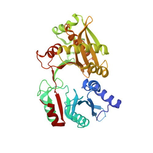
    Weissella viridescens FemX (FemX(Wv)) belongs to the Fem family of nonribosomal peptidyl transferases that use aminoacyl-tRNA as the amino acid donor to synthesize the peptide cross-bridge found in the peptidoglycan of many species of pathogenic gram-positive bacteria. We have recently solved the crystal structure of FemX(Wv) in complex with the peptidoglycan precursor UDP-MurNAc-pentapeptide and report here the site-directed mutagenesis of nine residues located in the binding cavity for this substrate. Two substitutions, Lys36Met and Arg211Met, depressed FemX(Wv) transferase activity below detectable levels without affecting protein folding. Analogues of UDP-MurNAc-pentapeptide lacking the phosphate groups or the C-terminal D-alanyl residues were not substrates of the enzyme. These results indicate that Lys36 and Arg211 participate in a complex hydrogen bond network that connects the C-terminal D-Ala residues to the phosphate groups of UDP-MurNAc-pentapeptide and constrains the substrate in a conformation that is essential for transferase activity.


  • Organizational Affiliation
    • Laboratoire de Recherche Moléculaire sur les Antibiotiques, INSERM U655, Université Paris 6, Paris, France.

Macromolecule Content 

  • Total Structure Weight: 38.22 kDa 
  • Atom Count: 3,054 
  • Modeled Residue Count: 335 
  • Deposited Residue Count: 335 
  • Unique protein chains: 1

Macromolecules

Find similar proteins by:|  3D Structure
Entity ID: 1
MoleculeChains  Sequence LengthOrganismDetailsImage
FemX335Weissella viridescensMutation(s): 1 
Gene Names: femx
EC: 2.3.2.10
UniProt
Find proteins for Q9EY50 (Weissella viridescens)
Explore Q9EY50 
Go to UniProtKB:  Q9EY50
Entity Groups
Sequence Clusters30% Identity50% Identity70% Identity90% Identity95% Identity100% Identity
UniProt GroupQ9EY50
Sequence Annotations
Expand
Reference Sequence

Small Molecules

Ligands 1 Unique
IDChains Name / Formula / InChI Key2D Diagram3D Interactions
MG

Query on MG


Download:Ideal Coordinates CCD File
B [auth A]MAGNESIUM ION
Mg
JLVVSXFLKOJNIY-UHFFFAOYSA-N

Experimental Data & Validation

Experimental Data

  • Method: X-RAY DIFFRACTION
  • Resolution: 1.95 Å
  • R-Value Free:  0.233 (Depositor), 0.233 (DCC) 
  • R-Value Work:  0.196 (Depositor), 0.196 (DCC) 
  • R-Value Observed: 0.196 (Depositor) 
Space Group: P 1 21 1
Unit Cell:
Length ( Å )Angle ( ˚ )
a = 42.326α = 90
b = 101.305β = 103.02
c = 46.331γ = 90
Software Package:
Software NamePurpose
CNSrefinement
MOSFLMdata reduction
CCP4data scaling
AMoREphasing

Structure Validation

View Full Validation Report


Entry History 

Deposition Data

Revision History  (Full details and data files)

  • Version 1.0: 2005-05-31
    Type: Initial release
  • Version 1.1: 2008-04-30
    Changes: Version format compliance
  • Version 1.2: 2011-07-13
    Changes: Version format compliance
  • Version 1.3: 2021-10-20
    Changes: Database references, Derived calculations
  • Version 1.4: 2023-08-23
    Changes: Data collection, Refinement description