1XDA | pdb_00001xda

STRUCTURE OF INSULIN


Experimental Data Snapshot

  • Method: X-RAY DIFFRACTION
  • Resolution: 1.80 Å
  • R-Value Work: 
    0.174 (Depositor) 

Starting Model: experimental
View more details

wwPDB Validation 3D Report Full Report

Validation slider image for 1XDA

This is version 1.4 of the entry. See complete history

Literature

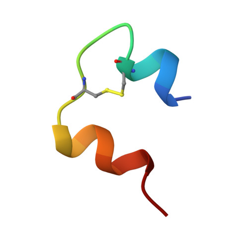
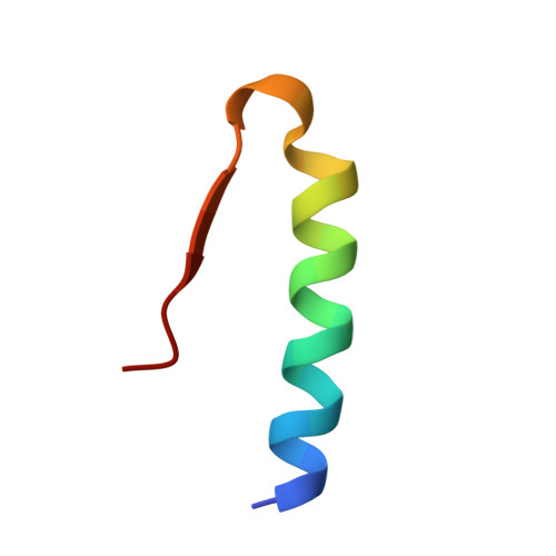
Crystal structure of a prolonged-acting insulin with albumin-binding properties.

Whittingham, J.L.Havelund, S.Jonassen, I.

(1997) Biochemistry 36: 2826-2831

  • DOI: https://doi.org/10.1021/bi9625105
  • Primary Citation Related Structures: 
    1XDA

  • PubMed Abstract: 

    The fatty acid acylated insulin, Lys(B29)-tetradecanoyl, des-(B30) human insulin, has been crystallized and the structure determined by X-ray crystallography. The fatty acid substituent on residue B29 Lys binds reversibly to circulating albumin protein in vivo, and by this mechanism the hormone's action is prolonged. Crystals of the fatty acid insulin grow in space group R3, with two dimers in the asymmetric unit, and diffract to 1.8 A spacing. The structure has been solved by molecular replacement and refined using a maximum likelihood method. The crystal structure consists of R6 zinc insulin hexamers which contain phenol. The fatty acids can be seen bound between the hexamers, making specific interactions with the side chains of residue B1 Phe; however, the lysine side chains to which the fatty acids are covalently attached are mostly disordered. The mode of binding of the fatty acids appears to be determined by crystal packing, and whether or not they interact with the protein in this way in solution remains uncertain.


  • Organizational Affiliation
    • Department of Chemistry, University of York, Heslington, England.

Macromolecule Content 

  • Total Structure Weight: 24.56 kDa 
  • Atom Count: 1,860 
  • Modeled Residue Count: 200 
  • Deposited Residue Count: 200 
  • Unique protein chains: 2

Macromolecules

Find similar proteins by:  Sequence   |   3D Structure  
Entity ID: 1
MoleculeChains  Sequence LengthOrganismDetailsImage
FATTY ACID ACYLATED INSULIN
A, C, E, G
21Homo sapiensMutation(s): 0 
UniProt & NIH Common Fund Data Resources
Find proteins for P01308 (Homo sapiens)
Explore P01308 
Go to UniProtKB:  P01308
PHAROS:  P01308
GTEx:  ENSG00000254647 
Entity Groups
Sequence Clusters30% Identity50% Identity70% Identity90% Identity95% Identity100% Identity
UniProt GroupP01308
Sequence Annotations
Expand
Reference Sequence
Find similar proteins by:|  3D Structure
Entity ID: 2
MoleculeChains  Sequence LengthOrganismDetailsImage
FATTY ACID ACYLATED INSULIN
B, D, F, H
29Homo sapiensMutation(s): 0 
UniProt & NIH Common Fund Data Resources
Find proteins for P01308 (Homo sapiens)
Explore P01308 
Go to UniProtKB:  P01308
PHAROS:  P01308
GTEx:  ENSG00000254647 
Entity Groups
Sequence Clusters30% Identity50% Identity70% Identity90% Identity95% Identity100% Identity
UniProt GroupP01308
Sequence Annotations
Expand
Reference Sequence

Small Molecules

Ligands 4 Unique
IDChains Name / Formula / InChI Key2D Diagram3D Interactions
MYR

Query on MYR


Download:Ideal Coordinates CCD File
L [auth B],
P [auth D],
T [auth F],
X [auth H]
MYRISTIC ACID
C14 H28 O2
TUNFSRHWOTWDNC-UHFFFAOYSA-N
IPH

Query on IPH


Download:Ideal Coordinates CCD File
I [auth A],
M [auth C],
Q [auth E],
U [auth G]
PHENOL
C6 H6 O
ISWSIDIOOBJBQZ-UHFFFAOYSA-N
ZN

Query on ZN


Download:Ideal Coordinates CCD File
J [auth B],
N [auth D],
R [auth F],
V [auth H]
ZINC ION
Zn
PTFCDOFLOPIGGS-UHFFFAOYSA-N
CL

Query on CL


Download:Ideal Coordinates CCD File
K [auth B],
O [auth D],
S [auth F],
W [auth H]
CHLORIDE ION
Cl
VEXZGXHMUGYJMC-UHFFFAOYSA-M

Experimental Data & Validation

Experimental Data

  • Method: X-RAY DIFFRACTION
  • Resolution: 1.80 Å
  • R-Value Work:  0.174 (Depositor) 
Space Group: H 3
Unit Cell:
Length ( Å )Angle ( ˚ )
a = 78.752α = 90
b = 78.752β = 90
c = 79.199γ = 120
Software Package:
Software NamePurpose
DENZOdata reduction
SCALEPACKdata scaling
AMoREphasing
REFMACrefinement

Structure Validation

View Full Validation Report


Entry History 

Deposition Data

Revision History  (Full details and data files)

  • Version 1.0: 1997-07-07
    Type: Initial release
  • Version 1.1: 2008-03-24
    Changes: Version format compliance
  • Version 1.2: 2011-07-13
    Changes: Version format compliance
  • Version 1.3: 2024-04-03
    Changes: Data collection, Database references, Derived calculations, Refinement description
  • Version 1.4: 2024-10-09
    Changes: Structure summary