1XAG | pdb_00001xag

CRYSTAL STRUCTURE OF STAPHLYOCOCCUS AUREUS 3-DEHYDROQUINATE SYNTHASE (DHQS) IN COMPLEX WITH ZN2+, NAD+ AND CARBAPHOSPHONATE


Experimental Data Snapshot

  • Method: X-RAY DIFFRACTION
  • Resolution: 2.45 Å
  • R-Value Free: 
    0.268 (Depositor), 0.269 (DCC) 
  • R-Value Work: 
    0.197 (Depositor), 0.197 (DCC) 
  • R-Value Observed: 
    0.197 (Depositor) 

Starting Model: experimental
View more details

wwPDB Validation 3D Report Full Report

Validation slider image for 1XAG

Ligand Structure Quality Assessment 


This is version 1.3 of the entry. See complete history

Literature

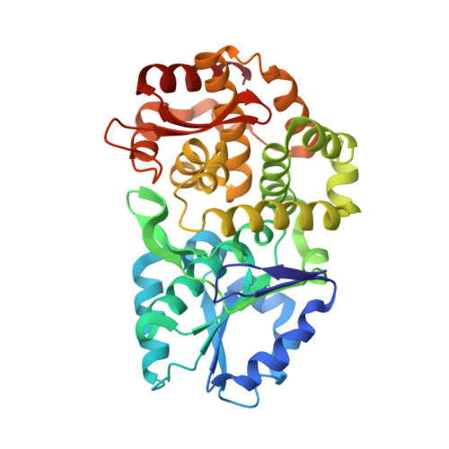
Comparison of ligand induced conformational changes and domain closure mechanisms, between prokaryotic and eukaryotic dehydroquinate synthases.

Nichols, C.E.Ren, J.Leslie, K.Dhaliwal, B.Lockyer, M.Charles, I.Hawkins, A.R.Stammers, D.K.

(2004) J Mol Biology 343: 533-546

  • DOI: https://doi.org/10.1016/j.jmb.2004.08.039
  • Primary Citation Related Structures: 
    1XAG, 1XAH, 1XAI, 1XAJ, 1XAL

  • PubMed Abstract: 

    Dehydroquinate synthase (DHQS) is a potential target for the development of novel broad-spectrum antimicrobial drugs, active against both prokaryotes and lower eukaryotes. Structures have been reported for Aspergillus nidulans DHQS (AnDHQS) in complexes with a range of ligands. Analysis of these AnDHQS structures showed that a large-scale domain movement occurs during the normal catalytic cycle, with a complex series of structural elements propagating substrate binding-induced conformational changes away from the active site to distal locations. Compared to corresponding fungal enzymes, DHQS from bacterial species are both mono-functional and significantly smaller. We have therefore determined the structure of Staphylococcus aureus DHQS (SaDHQS) in five liganded states, allowing comparison of ligand-induced conformational changes and mechanisms of domain closure between fungal and bacterial enzymes. This comparative analysis shows that substrate binding initiates a large-scale domain closure in both species' DHQS and that the active site stereochemistry, of the catalytically competent closed-form enzyme thus produced, is also highly conserved. However, comparison of AnDHQS and SaDHQS open-form structures, and analysis of the putative dynamic processes by which the transition to the closed-form states are made, shows a far lower degree of similarity, indicating a significant structural divergence. As a result, both the nature of the propagation of conformational change and the mechanical systems involved in this propagation are quite different between the DHQSs from the two species.


  • Organizational Affiliation
    • Division of Structural Biology, The Wellcome Trust Centre for Human Genetics, University of Oxford, Roosevelt Drive, Oxford OX3 7BN, UK.

Macromolecule Content 

  • Total Structure Weight: 41.54 kDa 
  • Atom Count: 3,020 
  • Modeled Residue Count: 353 
  • Deposited Residue Count: 354 
  • Unique protein chains: 1

Macromolecules

Find similar proteins by:|  3D Structure
Entity ID: 1
MoleculeChains  Sequence LengthOrganismDetailsImage
3-dehydroquinate synthase354Staphylococcus aureusMutation(s): 0 
Gene Names: aroB
EC: 4.2.3.4
UniProt
Find proteins for Q6GGU4 (Staphylococcus aureus (strain MRSA252))
Explore Q6GGU4 
Go to UniProtKB:  Q6GGU4
Entity Groups
Sequence Clusters30% Identity50% Identity70% Identity90% Identity95% Identity100% Identity
UniProt GroupQ6GGU4
Sequence Annotations
Expand
Reference Sequence

Small Molecules

Ligands 4 Unique
IDChains Name / Formula / InChI Key2D Diagram3D Interactions
NAD

Query on NAD


Download:Ideal Coordinates CCD File
H [auth A]NICOTINAMIDE-ADENINE-DINUCLEOTIDE
C21 H27 N7 O14 P2
BAWFJGJZGIEFAR-NNYOXOHSSA-N
CRB

Query on CRB


Download:Ideal Coordinates CCD File
I [auth A][1R-(1ALPHA,3BETA,4ALPHA,5BETA)]-5-(PHOSPHONOMETHYL)-1,3,4-TRIHYDROXYCYCLOHEXANE-1-CARBOXYLIC ACID
C8 H15 O8 P
BKLICLLAHMTUPK-UNGCPHIMSA-N
ZN

Query on ZN


Download:Ideal Coordinates CCD File
B [auth A]ZINC ION
Zn
PTFCDOFLOPIGGS-UHFFFAOYSA-N
CL

Query on CL


Download:Ideal Coordinates CCD File
C [auth A],
D [auth A],
E [auth A],
F [auth A],
G [auth A]
CHLORIDE ION
Cl
VEXZGXHMUGYJMC-UHFFFAOYSA-M

Experimental Data & Validation

Experimental Data

  • Method: X-RAY DIFFRACTION
  • Resolution: 2.45 Å
  • R-Value Free:  0.268 (Depositor), 0.269 (DCC) 
  • R-Value Work:  0.197 (Depositor), 0.197 (DCC) 
  • R-Value Observed: 0.197 (Depositor) 
Space Group: P 43 21 2
Unit Cell:
Length ( Å )Angle ( ˚ )
a = 53.29α = 90
b = 53.29β = 90
c = 240.07γ = 90
Software Package:
Software NamePurpose
DENZOdata reduction
SCALEPACKdata scaling
CNSrefinement
CNSphasing

Structure Validation

View Full Validation Report


Ligand Structure Quality Assessment 


Entry History 

Deposition Data

Revision History  (Full details and data files)

  • Version 1.0: 2005-03-01
    Type: Initial release
  • Version 1.1: 2008-04-30
    Changes: Version format compliance
  • Version 1.2: 2011-07-13
    Changes: Version format compliance
  • Version 1.3: 2023-08-23
    Changes: Data collection, Database references, Derived calculations, Refinement description