1X89 | pdb_00001x89

Crystal structure of Siderocalin (NGAL, Lipocalin 2) complexed with Carboxymycobactin S


Experimental Data Snapshot

  • Method: X-RAY DIFFRACTION
  • Resolution: 2.10 Å
  • R-Value Free: 
    0.251 (Depositor) 
  • R-Value Work: 
    0.220 (Depositor) 

Starting Model: experimental
View more details

wwPDB Validation 3D Report Full Report

Validation slider image for 1X89

This is version 1.6 of the entry. See complete history

Literature

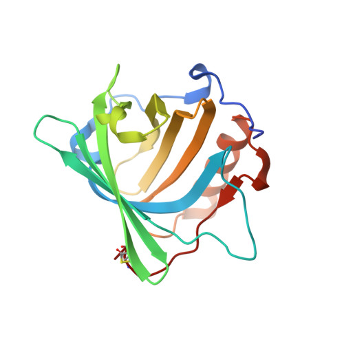
Siderocalin (Lcn 2) Also Binds Carboxymycobactins, Potentially Defending against Mycobacterial Infections through Iron Sequestration

Holmes, M.A.Paulsene, W.Jide, X.Ratledge, C.Strong, R.K.

(2005) Structure 13: 29-41

  • DOI: https://doi.org/10.1016/j.str.2004.10.009
  • Primary Citation Related Structures: 
    1X71, 1X89, 1X8U

  • PubMed Abstract: 

    Siderocalin, a member of the lipocalin family of binding proteins, is found in neutrophil granules, uterine secretions, and at markedly elevated levels in serum and synovium during bacterial infection; it is also secreted from epithelial cells in response to inflammation or tumorigenesis. Identification of high-affinity ligands, bacterial catecholate-type siderophores (such as enterochelin), suggested a possible function for siderocalin: an antibacterial agent, complementing the general antimicrobial innate immune system iron-depletion strategy, sequestering iron as ferric siderophore complexes. Supporting this hypothesis, siderocalin is a potent bacteriostatic agent in vitro under iron-limiting conditions and, when knocked out, renders mice remarkably susceptible to bacterial infection. Here we show that siderocalin also binds soluble siderophores of mycobacteria, including M. tuberculosis: carboxymycobactins. Siderocalin employs a degenerate recognition mechanism to cross react with these dissimilar types of siderophores, broadening the potential utility of this innate immune defense.


  • Organizational Affiliation
    • Division of Basic Sciences, Fred Hutchinson Cancer Research Center, 1100 Fairview Avenue North, Seattle, Washington 98109, USA.

Macromolecule Content 

  • Total Structure Weight: 64.07 kDa 
  • Atom Count: 4,408 
  • Modeled Residue Count: 518 
  • Deposited Residue Count: 534 
  • Unique protein chains: 1

Macromolecules

Find similar proteins by:|  3D Structure
Entity ID: 1
MoleculeChains  Sequence LengthOrganismDetailsImage
Neutrophil gelatinase-associated lipocalin
A, B, C
178Homo sapiensMutation(s): 1 
Gene Names: LCN2NGALHNL
UniProt & NIH Common Fund Data Resources
Find proteins for P80188 (Homo sapiens)
Explore P80188 
Go to UniProtKB:  P80188
PHAROS:  P80188
GTEx:  ENSG00000148346 
Entity Groups
Sequence Clusters30% Identity50% Identity70% Identity90% Identity95% Identity100% Identity
UniProt GroupP80188
Sequence Annotations
Expand
Reference Sequence

Experimental Data & Validation

Experimental Data

  • Method: X-RAY DIFFRACTION
  • Resolution: 2.10 Å
  • R-Value Free:  0.251 (Depositor) 
  • R-Value Work:  0.220 (Depositor) 
Space Group: P 41 21 2
Unit Cell:
Length ( Å )Angle ( ˚ )
a = 114.2α = 90
b = 114.2β = 90
c = 119.3γ = 90
Software Package:
Software NamePurpose
DENZOdata reduction
SCALEPACKdata scaling
CNSrefinement

Structure Validation

View Full Validation Report


Entry History 

Deposition Data

Revision History  (Full details and data files)

  • Version 1.0: 2005-01-25
    Type: Initial release
  • Version 1.1: 2008-04-30
    Changes: Version format compliance
  • Version 1.2: 2011-07-13
    Changes: Version format compliance
  • Version 1.3: 2017-10-11
    Changes: Refinement description
  • Version 1.4: 2021-10-20
    Changes: Database references, Derived calculations
  • Version 1.5: 2024-04-03
    Changes: Data collection, Refinement description
  • Version 1.6: 2024-11-06
    Changes: Structure summary