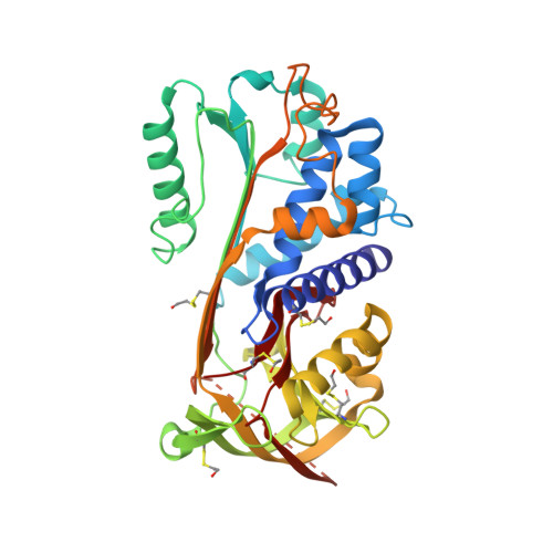
1WZ9

The 2.1 A structure of a tumour suppressing serpin


Experimental Data Snapshot

  • Method: X-RAY DIFFRACTION
  • Resolution: 2.10 Å
  • R-Value Free: 0.249 
  • R-Value Work: 0.205 
  • R-Value Observed: 0.207 

Starting Model: experimental
View more details

wwPDB Validation   3D Report Full Report


This is version 1.5 of the entry. See complete history


Literature

The high resolution crystal structure of the human tumor suppressor maspin reveals a novel conformational switch in the G-helix.

Law, R.H.Irving, J.A.Buckle, A.M.Ruzyla, K.Buzza, M.Bashtannyk-Puhalovich, T.A.Beddoe, T.C.Nguyen, K.Worrall, D.M.Bottomley, S.P.Bird, P.I.Rossjohn, J.Whisstock, J.C.

(2005) J Biol Chem 280: 22356-22364

  • DOI: https://doi.org/10.1074/jbc.M412043200
  • Primary Citation of Related Structures:  
    1WZ9, 1XU8

  • PubMed Abstract: 

    Maspin is a serpin that acts as a tumor suppressor in a range of human cancers, including tumors of the breast and lung. Maspin is crucial for development, because homozygous loss of the gene is lethal; however, the precise physiological role of the molecule is unclear. To gain insight into the function of human maspin, we have determined its crystal structure in two similar, but non-isomorphous crystal forms, to 2.1- and 2.8-A resolution, respectively. The structure reveals that maspin adopts the native serpin fold in which the reactive center loop is expelled fully from the A beta-sheet, makes minimal contacts with the core of the molecule, and exhibits a high degree of flexibility. A buried salt bridge unique to maspin orthologues causes an unusual bulge in the region around the D and E alpha-helices, an area of the molecule demonstrated in other serpins to be important for cofactor recognition. Strikingly, the structural data reveal that maspin is able to undergo conformational change in and around the G alpha-helix, switching between an open and a closed form. This change dictates the electrostatic character of a putative cofactor binding surface and highlights this region as a likely determinant of maspin function. The high resolution crystal structure of maspin provides a detailed molecular framework to elucidate the mechanism of function of this important tumor suppressor.


  • Organizational Affiliation

    The Protein Crystallography Unit, Monash Centre for Synchrotron Science and The Department of Biochemistry and Molecular Biology, School of Biomedical Sciences, Faculty of Medicine, Monash University, Clayton, Victoria 3800, Australia.


Macromolecules
Find similar proteins by:  (by identity cutoff)  |  3D Structure
Entity ID: 1
MoleculeChains Sequence LengthOrganismDetailsImage
Maspin precursor
A, B
375Homo sapiensMutation(s): 6 
Gene Names: SERPINB5
UniProt & NIH Common Fund Data Resources
Find proteins for P36952 (Homo sapiens)
Explore P36952 
Go to UniProtKB:  P36952
PHAROS:  P36952
GTEx:  ENSG00000206075 
Entity Groups  
Sequence Clusters30% Identity50% Identity70% Identity90% Identity95% Identity100% Identity
UniProt GroupP36952
Sequence Annotations
Expand
  • Reference Sequence
Small Molecules
Ligands 1 Unique
IDChains Name / Formula / InChI Key2D Diagram3D Interactions
SO4
Query on SO4

Download Ideal Coordinates CCD File 
C [auth A]SULFATE ION
O4 S
QAOWNCQODCNURD-UHFFFAOYSA-L
Modified Residues  1 Unique
IDChains TypeFormula2D DiagramParent
CME
Query on CME
A, B
L-PEPTIDE LINKINGC5 H11 N O3 S2CYS
Experimental Data & Validation

Experimental Data

  • Method: X-RAY DIFFRACTION
  • Resolution: 2.10 Å
  • R-Value Free: 0.249 
  • R-Value Work: 0.205 
  • R-Value Observed: 0.207 
  • Space Group: P 21 21 21
Unit Cell:
Length ( Å )Angle ( ˚ )
a = 54.115α = 90
b = 95.047β = 90
c = 139.225γ = 90
Software Package:
Software NamePurpose
REFMACrefinement
SCALEPACKdata scaling
AMoREphasing

Structure Validation

View Full Validation Report


Entry History 

Revision History  (Full details and data files)

  • Version 1.0: 2005-03-15
    Type: Initial release
  • Version 1.1: 2008-04-30
    Changes: Version format compliance
  • Version 1.2: 2011-07-13
    Changes: Version format compliance
  • Version 1.3: 2017-10-11
    Changes: Refinement description
  • Version 1.4: 2023-10-25
    Changes: Data collection, Database references, Derived calculations, Refinement description
  • Version 1.5: 2024-10-30
    Changes: Structure summary