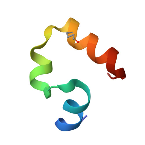
High-resolution x-ray crystal structures of the villin headpiece subdomain, an ultrafast folding protein.
Chiu, T.K., Kubelka, J., Herbst-Irmer, R., Eaton, W.A., Hofrichter, J., Davies, D.R.(2005) Proc Natl Acad Sci U S A 102: 7517-7522
- PubMed: 15894611
- DOI: https://doi.org/10.1073/pnas.0502495102
- Primary Citation of Related Structures:
1WY3, 1WY4, 1YRF, 1YRI - PubMed Abstract:
The 35-residue subdomain of the villin headpiece (HP35) is a small ultrafast folding protein that is being intensely studied by experiments, theory, and simulations. We have solved the x-ray structures of HP35 and its fastest folding mutant [K24 norleucine (nL)] to atomic resolution and compared their experimentally measured folding kinetics by using laser temperature jump. The structures, which are in different space groups, are almost identical to each other but differ significantly from previously solved NMR structures. Hence, the differences between the x-ray and NMR structures are probably not caused by lattice contacts or crystal/solution differences, but reflect the higher accuracy of the x-ray structures. The x-ray structures reveal important details of packing of the hydrophobic core and some additional features, such as cross-helical H bonds. Comparison of the x-ray structures indicates that the nL substitution produces only local perturbations. Consequently, the finding that the small stabilization by the mutation is completely reflected in an increased folding rate suggests that this region of the protein is as structured in the transition state as in the folded structure. It is therefore a target for engineering to increase the folding rate of the subdomain from approximately 0.5 micros(-1) for the nL mutant to the estimated theoretical speed limit of approximately 3 micros(-1).
Organizational Affiliation:
Laboratory of Molecular Biology, National Institute of Diabetes and Digestive and Kidney Diseases, National Institutes of Health, Bethesda, MD 20892-0520, USA.


