1WVH | pdb_00001wvh

Crystal structure of tensin1 PTB domain


Experimental Data Snapshot

  • Method: X-RAY DIFFRACTION
  • Resolution: 1.50 Å
  • R-Value Free: 
    0.236 (Depositor), 0.235 (DCC) 
  • R-Value Work: 
    0.218 (Depositor), 0.218 (DCC) 
  • R-Value Observed: 
    0.218 (Depositor) 

wwPDB Validation 3D Report Full Report

Validation slider image for 1WVH

This is version 1.3 of the entry. See complete history

Literature

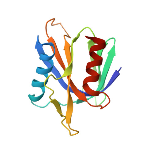
Structure of the PTB domain of tensin1 and a model for its recruitment to fibrillar adhesions.

McCleverty, C.J.Lin, D.C.Liddington, R.C.

(2007) Protein Sci 16: 1223-1229

  • DOI: https://doi.org/10.1110/ps.072798707
  • Primary Citation Related Structures: 
    1WVH

  • PubMed Abstract: 

    Tensin is a cytoskeletal protein that links integrins to the actin cytoskeleton at sites of cell-matrix adhesion. Here we describe the crystal structure of the phosphotyrosine-binding (PTB) domain of tensin1, and show that it binds integrins in an NPxY-dependent fashion. Alanine mutagenesis of both the PTB domain and integrin tails supports a model of integrin binding similar to that of the PTB-like domain of talin. However, we also show that phosphorylation of the NPxY tyrosine, which disrupts talin binding, has a negligible effect on tensin binding. This suggests that tyrosine phosphorylation of integrin, which occurs during the maturation of focal adhesions, could act as a switch to promote the migration of tensin-integrin complexes into fibronectin-mediated fibrillar adhesions.


  • Organizational Affiliation
    • Cancer Center, Burnham Institute for Medical Research, La Jolla, California 92037, USA.

Macromolecule Content 

  • Total Structure Weight: 14.64 kDa 
  • Atom Count: 1,120 
  • Modeled Residue Count: 132 
  • Deposited Residue Count: 134 
  • Unique protein chains: 1

Macromolecules

Find similar proteins by:|  3D Structure
Entity ID: 1
MoleculeChains  Sequence LengthOrganismDetailsImage
Tensin134Gallus gallusMutation(s): 0 
EC: 3.1.3
UniProt
Find proteins for Q04205 (Gallus gallus)
Explore Q04205 
Go to UniProtKB:  Q04205
Entity Groups
Sequence Clusters30% Identity50% Identity70% Identity90% Identity95% Identity100% Identity
UniProt GroupQ04205
Sequence Annotations
Expand
Reference Sequence

Experimental Data & Validation

Experimental Data

  • Method: X-RAY DIFFRACTION
  • Resolution: 1.50 Å
  • R-Value Free:  0.236 (Depositor), 0.235 (DCC) 
  • R-Value Work:  0.218 (Depositor), 0.218 (DCC) 
  • R-Value Observed: 0.218 (Depositor) 
Space Group: I 4
Unit Cell:
Length ( Å )Angle ( ˚ )
a = 75.68α = 90
b = 75.68β = 90
c = 48.69γ = 90
Software Package:
Software NamePurpose
DENZOdata reduction
SCALEPACKdata scaling
SOLVEphasing
CNSrefinement

Structure Validation

View Full Validation Report


Entry History 

Deposition Data

Revision History  (Full details and data files)

  • Version 1.0: 2005-12-13
    Type: Initial release
  • Version 1.1: 2008-04-30
    Changes: Version format compliance
  • Version 1.2: 2011-07-13
    Changes: Version format compliance
  • Version 1.3: 2024-03-13
    Changes: Data collection, Database references