


1WSI | pdb_00001wsi
1WSI | pdb_00001wsi
Crystal structure of E.coli RNase HI active site mutant (E48A/K87A/D134N)
- PDB DOI: https://doi.org/10.2210/pdb1WSI/pdb
- Classification: HYDROLASE
- Organism(s): Escherichia coli
- Expression System: Escherichia coli
- Mutation(s): Yes
- Deposited: 2004-11-07 Released: 2004-11-23
Experimental Data Snapshot
- Method: X-RAY DIFFRACTION
- Resolution: 2.00 Å
- R-Value Free: 0.258 (Depositor)
- R-Value Work: 0.217 (Depositor)
wwPDB Validation 3D Report Full Report

This is version 1.4 of the entry. See complete history.

Macromolecules
Find similar proteins by:
| 3D Structure
Entity ID: 1 | |||||
|---|---|---|---|---|---|
| Molecule | Chains | Sequence Length | Organism | Details | Image |
| Ribonuclease HI | 155 | Escherichia coli | Mutation(s): 3 EC: 3.1.26.4 | 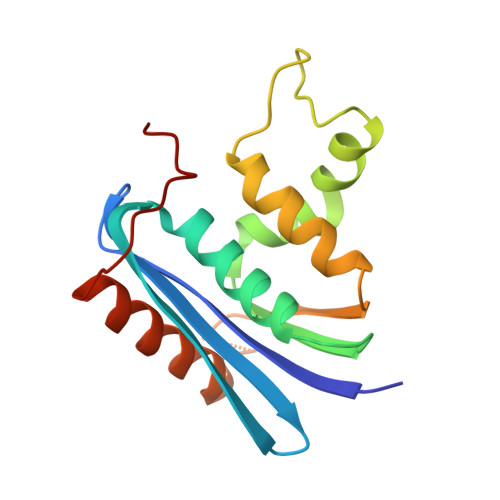 | |
UniProt | |||||
Entity Groups | |||||
| Sequence Clusters | 30% Identity50% Identity70% Identity90% Identity95% Identity100% Identity | ||||
| UniProt Group | P0A7Y4 | ||||
Sequence AnnotationsExpand | |||||
Reference Sequence | |||||
Experimental Data & Validation
Experimental Data
- Method: X-RAY DIFFRACTION
- Resolution: 2.00 Å
- R-Value Free: 0.258 (Depositor)
- R-Value Work: 0.217 (Depositor)
Space Group: P 21 21 2
Unit Cell:
| Length ( Å ) | Angle ( ˚ ) |
|---|---|
| a = 121.12 | α = 90 |
| b = 80.12 | β = 90 |
| c = 64.458 | γ = 90 |
| Software Name | Purpose |
|---|---|
| CNS | refinement |
Entry History
Deposition Data
- Released Date: 2004-11-23 Deposition Author(s): Tsunaka, Y., Takano, K., Matsumura, H., Yamagata, Y., Kanaya, S.
Revision History (Full details and data files)
- Version 1.0: 2004-11-23
Type: Initial release - Version 1.1: 2008-04-30
Changes: Version format compliance - Version 1.2: 2011-07-13
Changes: Version format compliance - Version 1.3: 2021-11-10
Changes: Database references - Version 1.4: 2024-05-29
Changes: Data collection


