1WQO

CONTRIBUTION OF HYDROGEN BONDS TO THE CONFORMATIONAL STABILITY OF HUMAN LYSOZYME


Experimental Data Snapshot

  • Method: X-RAY DIFFRACTION
  • Resolution: 1.80 Å
  • R-Value Work: 0.170 
  • R-Value Observed: 0.170 

Starting Model: experimental
View more details

wwPDB Validation   3D Report Full Report


This is version 1.4 of the entry. See complete history


Literature

Contribution of hydrogen bonds to the conformational stability of human lysozyme: calorimetry and X-ray analysis of six tyrosine --> phenylalanine mutants.

Yamagata, Y.Kubota, M.Sumikawa, Y.Funahashi, J.Takano, K.Fujii, S.Yutani, K.

(1998) Biochemistry 37: 9355-9362

  • DOI: https://doi.org/10.1021/bi980431i
  • Primary Citation of Related Structures:  
    1WQM, 1WQN, 1WQO, 1WQP, 1WQQ, 1WQR

  • PubMed Abstract: 

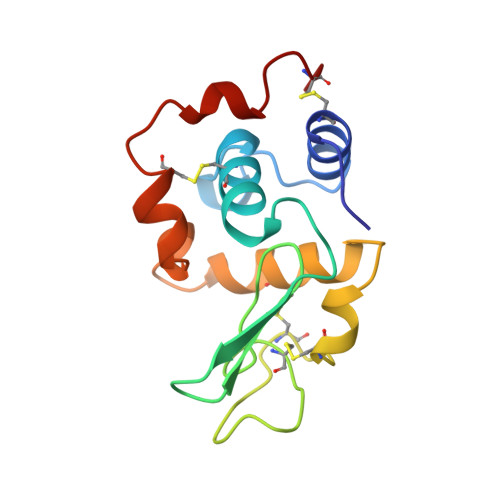
    The contribution of hydrogen bonds to the conformational stability of human lysozyme was investigated by the combination of calorimetric and X-ray analyses of six Tyr --> Phe mutants. Unfolding Delta G and unfolding Delta H values of the Tyr --> Phe mutant proteins were changed by from +0.3 to -4.0 kJ/mol and from 0 to -16 kJ/mol, respectively, compared to those of the wild-type protein. The net contribution of a hydrogen bond at a specific site to stability (Delta Gwild/HB), considering factors affected by substitutions, was evaluated on the basis of X-ray structures of the mutant proteins. In the present study, one of six mutant proteins was suitable for evaluating the strength of the hydrogen bond. Delta Gwild/HB for the intramolecular hydrogen bond at Tyr124 was evaluated to be 7.5 kJ/mol. Results of the analysis of other mutants also suggest that hydrogen bonds of the hydroxyl group of Tyr, including the hydrogen bond with a water molecule, contribute to the stabilization of the human lysozyme.


  • Organizational Affiliation

    Institute for Protein Research, Faculty of Pharmaceutical Sciences, Osaka University, Japan.


Macromolecules
Find similar proteins by:  (by identity cutoff)  |  3D Structure
Entity ID: 1
MoleculeChains Sequence LengthOrganismDetailsImage
LYSOZYME130Homo sapiensMutation(s): 1 
EC: 3.2.1.17
UniProt & NIH Common Fund Data Resources
Find proteins for P61626 (Homo sapiens)
Explore P61626 
Go to UniProtKB:  P61626
GTEx:  ENSG00000090382 
Entity Groups  
Sequence Clusters30% Identity50% Identity70% Identity90% Identity95% Identity100% Identity
UniProt GroupP61626
Sequence Annotations
Expand
  • Reference Sequence
Small Molecules
Ligands 2 Unique
IDChains Name / Formula / InChI Key2D Diagram3D Interactions
CL
Query on CL

Download Ideal Coordinates CCD File 
C [auth A],
D [auth A]
CHLORIDE ION
Cl
VEXZGXHMUGYJMC-UHFFFAOYSA-M
NA
Query on NA

Download Ideal Coordinates CCD File 
B [auth A]SODIUM ION
Na
FKNQFGJONOIPTF-UHFFFAOYSA-N
Experimental Data & Validation

Experimental Data

  • Method: X-RAY DIFFRACTION
  • Resolution: 1.80 Å
  • R-Value Work: 0.170 
  • R-Value Observed: 0.170 
  • Space Group: P 21 21 21
Unit Cell:
Length ( Å )Angle ( ˚ )
a = 56.65α = 90
b = 61.02β = 90
c = 33.74γ = 90
Software Package:
Software NamePurpose
PROCESSdata collection
PROCESSdata reduction
X-PLORmodel building
X-PLORrefinement
PROCESSdata scaling
X-PLORphasing

Structure Validation

View Full Validation Report


Entry History 

Deposition Data

Revision History  (Full details and data files)

  • Version 1.0: 1998-07-01
    Type: Initial release
  • Version 1.1: 2008-03-24
    Changes: Version format compliance
  • Version 1.2: 2011-07-13
    Changes: Version format compliance
  • Version 1.3: 2021-11-03
    Changes: Database references, Derived calculations, Other
  • Version 1.4: 2024-04-03
    Changes: Data collection, Refinement description