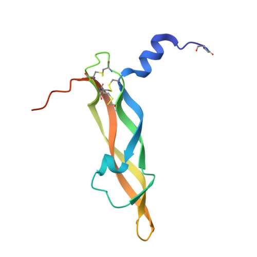
1WQ8

Crystal structure of Vammin, a VEGF-F from a snake venom


Experimental Data Snapshot

  • Method: X-RAY DIFFRACTION
  • Resolution: 1.90 Å
  • R-Value Free: 0.233 
  • R-Value Work: 0.192 
  • R-Value Observed: 0.192 

Starting Model: experimental
View more details

wwPDB Validation   3D Report Full Report


This is version 2.1 of the entry. See complete history


Literature

Crystal structures of novel vascular endothelial growth factors (VEGF) from snake venoms: insight into selective VEGF binding to kinase insert domain-containing receptor but not to fms-like tyrosine kinase-1.

Suto, K.Yamazaki, Y.Morita, T.Mizuno, H.

(2005) J Biol Chem 280: 2126-2131

  • DOI: https://doi.org/10.1074/jbc.M411395200
  • Primary Citation of Related Structures:  
    1WQ8, 1WQ9

  • PubMed Abstract: 

    Vascular endothelial growth factor-A (VEGF-A(165)) exerts multiple effects upon binding to the fms-like tyrosine kinase-1 (Flt-1) and the kinase insert domain-containing receptor (KDR). We recently identified two novel snake venom VEGFs (vammin and VR-1) having unique properties. These VEGFs, designated VEGF-Fs, are highly specific ligands for the kinase insert domain-containing receptor and exhibit potent biological activity both in vitro and in vivo when compared with VEGF-A(165). Here, we solved the crystal structures of vammin and VR-1 at 1.9 and 2.0 A resolutions, respectively. Both structures are very similar to each other, and these structures exhibit similar but significantly different features from the known structures of other VEGFs. These differences include a conformational difference in receptor-binding loop 3 caused by an amino acid residue insertion and a difference in surface potential on the possible binding surface for domain 3 of the receptor. These structural differences may be related to the highly selective ligand properties of VEGF-F.


  • Organizational Affiliation

    Department of Biochemistry, National Institute of Agrobiological Sciences, 2-1-2, Kannondai, Tsukuba, Ibaraki 305-8602, Japan.


Macromolecules
Find similar proteins by:  (by identity cutoff)  |  3D Structure
Entity ID: 1
MoleculeChains Sequence LengthOrganismDetailsImage
Vascular endothelial growth factor toxin110Vipera aspis aspisMutation(s): 0 
UniProt
Find proteins for P67863 (Vipera ammodytes ammodytes)
Explore P67863 
Go to UniProtKB:  P67863
Entity Groups  
Sequence Clusters30% Identity50% Identity70% Identity90% Identity95% Identity100% Identity
UniProt GroupP67863
Sequence Annotations
Expand
  • Reference Sequence
Small Molecules
Ligands 1 Unique
IDChains Name / Formula / InChI Key2D Diagram3D Interactions
TRS
Query on TRS

Download Ideal Coordinates CCD File 
B [auth A]2-AMINO-2-HYDROXYMETHYL-PROPANE-1,3-DIOL
C4 H12 N O3
LENZDBCJOHFCAS-UHFFFAOYSA-O
Modified Residues  1 Unique
IDChains TypeFormula2D DiagramParent
PCA
Query on PCA
A
L-PEPTIDE LINKINGC5 H7 N O3GLN
Experimental Data & Validation

Experimental Data

  • Method: X-RAY DIFFRACTION
  • Resolution: 1.90 Å
  • R-Value Free: 0.233 
  • R-Value Work: 0.192 
  • R-Value Observed: 0.192 
  • Space Group: I 4 2 2
Unit Cell:
Length ( Å )Angle ( ˚ )
a = 85.526α = 90
b = 85.526β = 90
c = 86.365γ = 90
Software Package:
Software NamePurpose
CNSrefinement
HKL-2000data reduction
SCALEPACKdata scaling
CNSphasing

Structure Validation

View Full Validation Report


Entry History 

Deposition Data

Revision History  (Full details and data files)

  • Version 1.0: 2004-12-07
    Type: Initial release
  • Version 1.1: 2008-04-30
    Changes: Version format compliance
  • Version 1.2: 2011-07-13
    Changes: Derived calculations, Version format compliance
  • Version 2.0: 2019-12-25
    Changes: Derived calculations, Polymer sequence
  • Version 2.1: 2024-04-03
    Changes: Data collection, Database references, Derived calculations, Refinement description