1WPL | pdb_00001wpl

Crystal structure of the inhibitory form of rat GTP cyclohydrolase I/GFRP complex


Experimental Data Snapshot

  • Method: X-RAY DIFFRACTION
  • Resolution: 2.80 Å
  • R-Value Free: 
    0.233 (Depositor), 0.234 (DCC) 
  • R-Value Work: 
    0.207 (Depositor), 0.205 (DCC) 

Starting Model: experimental
View more details

wwPDB Validation 3D Report Full Report

Validation slider image for 1WPL

Ligand Structure Quality Assessment 


This is version 1.3 of the entry. See complete history

Literature

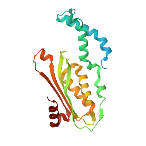
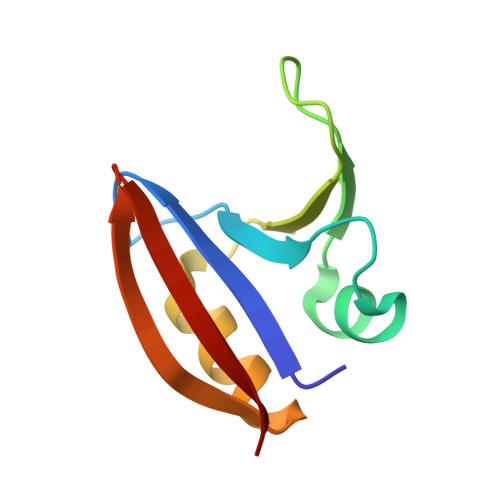
Structural basis of biopterin-induced inhibition of GTP cyclohydrolase I by GFRP, its feedback regulatory protein

Maita, N.Hatakeyama, K.Okada, K.Hakoshima, T.

(2004) J Biological Chem 279: 51534-51540

  • DOI: https://doi.org/10.1074/jbc.M409440200
  • Primary Citation Related Structures: 
    1WPL

  • PubMed Abstract: 

    GTP cyclohydrolase I (GTPCHI) is the rate-limiting enzyme involved in the biosynthesis of tetrahydrobiopterin, a key cofactor necessary for nitric oxide synthase and for the hydroxylases that are involved in the production of catecholamines and serotonin. In animals, the GTPCHI feedback regulatory protein (GFRP) binds GTPCHI to mediate feed-forward activation of GTPCHI activity in the presence of phenylalanine, whereas it induces feedback inhibition of enzyme activity in the presence of biopterin. Here, we have reported the crystal structure of the biopterin-induced inhibitory complex of GTPCHI and GFRP and compared it with the previously reported phenylalanine-induced stimulatory complex. The structure reveals five biopterin molecules located at each interface between GTPCHI and GFRP. Induced fitting structural changes by the biopterin binding expand large conformational changes in GTPCHI peptide segments forming the active site, resulting in inhibition of the activity. By locating 3,4-dihydroxy-phenylalanine-responsive dystonia mutations in the complex structure, we found mutations that may possibly disturb the GFRP-mediated regulation of GTPCHI.


  • Organizational Affiliation
    • Structural Biology Laboratory, Nara Institute of Science and Technology, CREST, Japan Science and Technology Agency, 8916-5 Takayama, Ikoma, Nara 630-0192, Japan.

Macromolecule Content 

  • Total Structure Weight: 360.88 kDa 
  • Atom Count: 22,650 
  • Modeled Residue Count: 2,780 
  • Deposited Residue Count: 3,140 
  • Unique protein chains: 2

Macromolecules

Find similar proteins by:|  3D Structure
Entity ID: 1
MoleculeChains  Sequence LengthOrganismDetailsImage
GTP cyclohydrolase I
A, B, C, D, E
A, B, C, D, E, F, G, H, I, J
230Rattus norvegicusMutation(s): 0 
EC: 3.5.4.16
UniProt
Find proteins for P22288 (Rattus norvegicus)
Explore P22288 
Go to UniProtKB:  P22288
Entity Groups
Sequence Clusters30% Identity50% Identity70% Identity90% Identity95% Identity100% Identity
UniProt GroupP22288
Sequence Annotations
Expand
Reference Sequence
Find similar proteins by:|  3D Structure
Entity ID: 2
MoleculeChains  Sequence LengthOrganismDetailsImage
GTP cyclohydrolase I feedback regulatory protein
K, L, M, N, O
K, L, M, N, O, P, Q, R, S, T
84Rattus norvegicusMutation(s): 0 
UniProt
Find proteins for P70552 (Rattus norvegicus)
Explore P70552 
Go to UniProtKB:  P70552
Entity Groups
Sequence Clusters30% Identity50% Identity70% Identity90% Identity95% Identity100% Identity
UniProt GroupP70552
Sequence Annotations
Expand
Reference Sequence

Small Molecules

Ligands 4 Unique
IDChains Name / Formula / InChI Key2D Diagram3D Interactions
3PO

Query on 3PO


Download:Ideal Coordinates CCD File
AA [auth B]
DA [auth C]
GA [auth D]
IA [auth E]
MA [auth F]
AA [auth B],
DA [auth C],
GA [auth D],
IA [auth E],
MA [auth F],
PA [auth G],
SA [auth H],
VA [auth I],
X [auth A],
XA [auth J]
TRIPHOSPHATE
H5 O10 P3
UNXRWKVEANCORM-UHFFFAOYSA-N
HBI

Query on HBI


Download:Ideal Coordinates CCD File
CA [auth C]
FA [auth D]
KA [auth F]
LA [auth F]
OA [auth G]
CA [auth C],
FA [auth D],
KA [auth F],
LA [auth F],
OA [auth G],
RA [auth H],
UA [auth I],
V [auth A],
W [auth A],
Z [auth B]
7,8-DIHYDROBIOPTERIN
C9 H13 N5 O3
FEMXZDUTFRTWPE-DZSWIPIPSA-N
ZN

Query on ZN


Download:Ideal Coordinates CCD File
BA [auth C]
EA [auth D]
HA [auth E]
JA [auth F]
NA [auth G]
BA [auth C],
EA [auth D],
HA [auth E],
JA [auth F],
NA [auth G],
QA [auth H],
TA [auth I],
U [auth A],
WA [auth J],
Y [auth B]
ZINC ION
Zn
PTFCDOFLOPIGGS-UHFFFAOYSA-N
NA

Query on NA


Download:Ideal Coordinates CCD File
AB [auth M]
BB [auth N]
CB [auth O]
DB [auth P]
EB [auth Q]
AB [auth M],
BB [auth N],
CB [auth O],
DB [auth P],
EB [auth Q],
FB [auth R],
GB [auth S],
HB [auth T],
YA [auth K],
ZA [auth L]
SODIUM ION
Na
FKNQFGJONOIPTF-UHFFFAOYSA-N

Experimental Data & Validation

Experimental Data

  • Method: X-RAY DIFFRACTION
  • Resolution: 2.80 Å
  • R-Value Free:  0.233 (Depositor), 0.234 (DCC) 
  • R-Value Work:  0.207 (Depositor), 0.205 (DCC) 
Space Group: P 1 21 1
Unit Cell:
Length ( Å )Angle ( ˚ )
a = 121.72α = 90
b = 109.67β = 97.85
c = 130.27γ = 90
Software Package:
Software NamePurpose
MOSFLMdata reduction
SCALAdata scaling
AMoREphasing
CNSrefinement
CCP4data scaling

Structure Validation

View Full Validation Report


Ligand Structure Quality Assessment 


Entry History 

Deposition Data

Revision History  (Full details and data files)

  • Version 1.0: 2004-09-28
    Type: Initial release
  • Version 1.1: 2008-04-30
    Changes: Version format compliance
  • Version 1.2: 2011-07-13
    Changes: Version format compliance
  • Version 1.3: 2023-10-25
    Changes: Data collection, Database references, Derived calculations, Refinement description