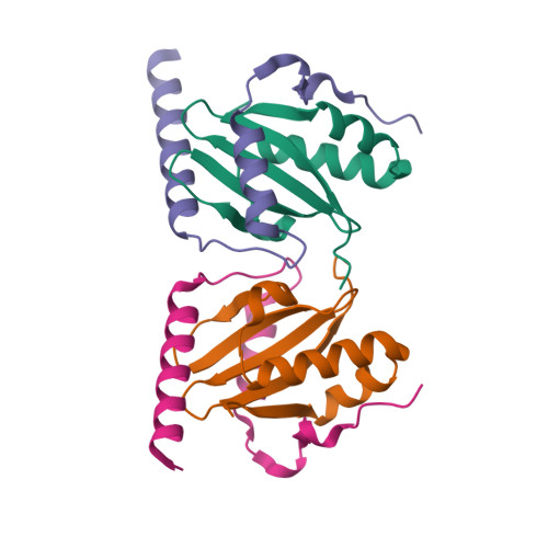
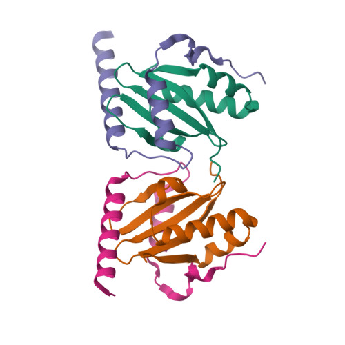
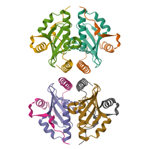
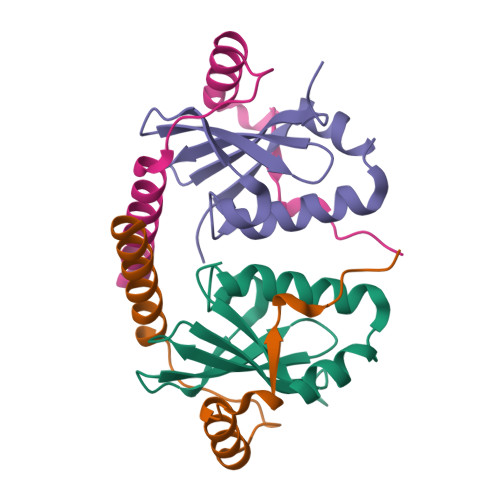
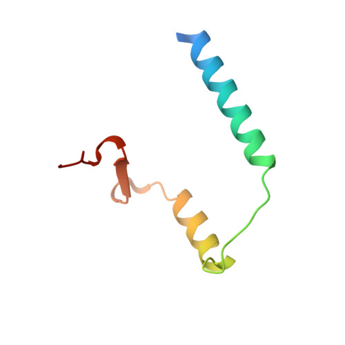
1WMI

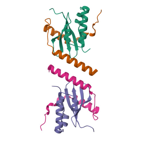
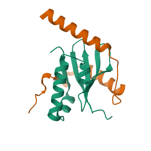
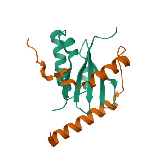
Crystal structure of archaeal RelE-RelB complex from Pyrococcus horikoshii OT3


Experimental Data Snapshot

  • Method: X-RAY DIFFRACTION
  • Resolution: 2.30 Å
  • R-Value Free: 0.276 
  • R-Value Work: 0.228 

wwPDB Validation   3D Report Full Report


This is version 1.3 of the entry. See complete history


Literature

Crystal structure of archaeal toxin-antitoxin RelE-RelB complex with implications for toxin activity and antitoxin effects

Takagi, H.Kakuta, Y.Okada, T.Yao, M.Tanaka, I.Kimura, M.

(2005) Nat Struct Mol Biol 12: 327-331

  • DOI: https://doi.org/10.1038/nsmb911
  • Primary Citation of Related Structures:  
    1WMI

  • PubMed Abstract: 

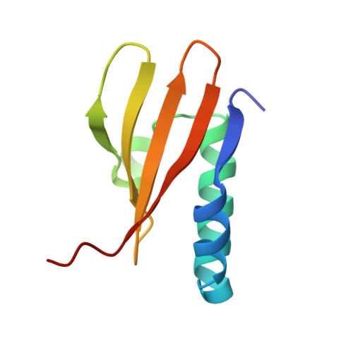
    The Escherichia coli chromosome encodes toxin-antitoxin pairs. The toxin RelE cleaves mRNA positioned at the A-site in ribosomes, whereas the antitoxin RelB relieves the effect of RelE. The hyperthermophilic archaeon Pyrococcus horikoshii OT3 has the archaeal homologs aRelE and aRelB. Here we report the crystal structure of aRelE in complex with aRelB determined at a resolution of 2.3 A. aRelE folds into an alpha/beta structure, whereas aRelB lacks a distinct hydrophobic core and extensively wraps around the molecular surface of aRelE. Neither component shows structural homology to known ribonucleases or their inhibitors. Site-directed mutagenesis suggests that Arg85, in the C-terminal region, is strongly involved in the functional activity of aRelE, whereas Arg40, Leu48, Arg58 and Arg65 play a modest role in the toxin's activity.


  • Organizational Affiliation

    Laboratory of Biochemistry, Department of Bioscience and Biotechnology, Faculty of Agriculture, Graduate School, Kyushu University, Fukuoka 812-8581, Japan.


Macromolecules
Find similar proteins by:  (by identity cutoff)  |  3D Structure
Entity ID: 1
MoleculeChains Sequence LengthOrganismDetailsImage
hypothetical protein PHS013
A, C
90Pyrococcus horikoshii OT3Mutation(s): 0 
UniProt
Find proteins for O73966 (Pyrococcus horikoshii (strain ATCC 700860 / DSM 12428 / JCM 9974 / NBRC 100139 / OT-3))
Explore O73966 
Go to UniProtKB:  O73966
Entity Groups  
Sequence Clusters30% Identity50% Identity70% Identity90% Identity95% Identity100% Identity
UniProt GroupO73966
Sequence Annotations
Expand
  • Reference Sequence
Find similar proteins by:  (by identity cutoff)  |  3D Structure
Entity ID: 2
MoleculeChains Sequence LengthOrganismDetailsImage
hypothetical protein PHS014
B, D
67Pyrococcus horikoshii OT3Mutation(s): 0 
UniProt
Find proteins for O73967 (Pyrococcus horikoshii (strain ATCC 700860 / DSM 12428 / JCM 9974 / NBRC 100139 / OT-3))
Explore O73967 
Go to UniProtKB:  O73967
Entity Groups  
Sequence Clusters30% Identity50% Identity70% Identity90% Identity95% Identity100% Identity
UniProt GroupO73967
Sequence Annotations
Expand
  • Reference Sequence
Experimental Data & Validation

Experimental Data

  • Method: X-RAY DIFFRACTION
  • Resolution: 2.30 Å
  • R-Value Free: 0.276 
  • R-Value Work: 0.228 
  • Space Group: P 21 21 2
Unit Cell:
Length ( Å )Angle ( ˚ )
a = 47.533α = 90
b = 142.651β = 90
c = 37.168γ = 90
Software Package:
Software NamePurpose
SCALEPACKdata scaling
SOLVEphasing
CNSrefinement
DENZOdata reduction

Structure Validation

View Full Validation Report


Entry History 

Deposition Data

Revision History  (Full details and data files)

  • Version 1.0: 2005-03-15
    Type: Initial release
  • Version 1.1: 2008-04-30
    Changes: Version format compliance
  • Version 1.2: 2011-07-13
    Changes: Derived calculations, Source and taxonomy, Version format compliance
  • Version 1.3: 2024-03-13
    Changes: Data collection, Database references