1WFD | pdb_00001wfd

Solution structure of mouse MIT domain


Experimental Data Snapshot

  • Method: SOLUTION NMR
  • Conformers Calculated: 100 
  • Conformers Submitted: 20 
  • Selection Criteria: structures with the least restraint violations, target function 

wwPDB Validation 3D Report Full Report

Validation slider image for 1WFD

This is version 1.4 of the entry. See complete history

Literature

Solution structure of mouse MIT domain

Suetake, T.Hayashi, F.Yokoyama, S.

To be published.

Macromolecule Content 

  • Total Structure Weight: 10.06 kDa 
  • Atom Count: 700 
  • Modeled Residue Count: 93 
  • Deposited Residue Count: 93 
  • Unique protein chains: 1

Macromolecules

Find similar proteins by:|  3D Structure
Entity ID: 1
MoleculeChains  Sequence LengthOrganismDetailsImage
Hypothetical protein 1500032H1893Mus musculusMutation(s): 0 
Gene Names: RIKEN cDNA 1500032H18
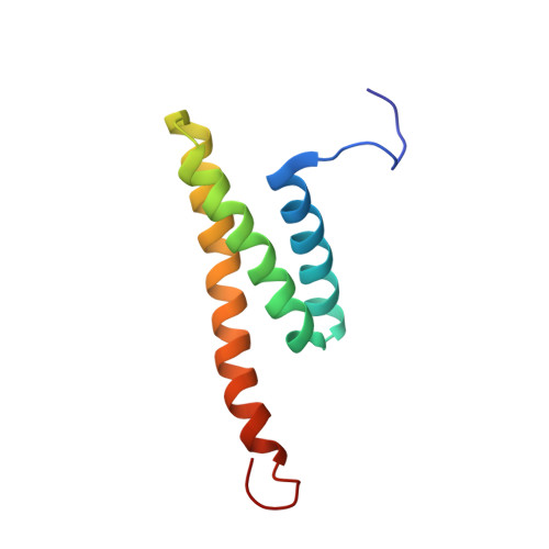
UniProt & NIH Common Fund Data Resources
Find proteins for Q8VDV8 (Mus musculus)
Explore Q8VDV8 
Go to UniProtKB:  Q8VDV8
IMPC:  MGI:1916278
Entity Groups
Sequence Clusters30% Identity50% Identity70% Identity90% Identity95% Identity100% Identity
UniProt GroupQ8VDV8
Sequence Annotations
Expand
Reference Sequence

Experimental Data & Validation

Experimental Data

  • Method: SOLUTION NMR
  • Conformers Calculated: 100 
  • Conformers Submitted: 20 
  • Selection Criteria: structures with the least restraint violations, target function 

Structure Validation

View Full Validation Report


Entry History 

Deposition Data

Revision History  (Full details and data files)

  • Version 1.0: 2004-11-26
    Type: Initial release
  • Version 1.1: 2008-04-30
    Changes: Version format compliance
  • Version 1.2: 2011-07-13
    Changes: Version format compliance
  • Version 1.3: 2022-03-02
    Changes: Data collection, Database references, Derived calculations
  • Version 1.4: 2024-05-29
    Changes: Data collection