1WCK | pdb_00001wck

Crystal structure of the C-terminal domain of BclA, the major antigen of the exosporium of the Bacillus anthracis spore.


Experimental Data Snapshot

  • Method: X-RAY DIFFRACTION
  • Resolution: 1.36 Å
  • R-Value Free: 
    0.190 (Depositor), 0.188 (DCC) 
  • R-Value Work: 
    0.179 (Depositor) 
  • R-Value Observed: 
    0.179 (Depositor) 

wwPDB Validation 3D Report Full Report

Validation slider image for 1WCK

This is version 1.4 of the entry. See complete history

Literature

The Crystal Structure of the Bacillus Anthracis Spore Surface Protein Bcla Shows Remarkable Similarity to Mammalian Proteins.

Rety, S.Salamitou, S.Garcia-Verdugo, I.Hulmes, D.J.Lehegarat, F.Chaby, R.Lewit-Bentley, A.

(2005) J Biological Chem 280: 43073

  • DOI: https://doi.org/10.1074/jbc.M510087200
  • Primary Citation Related Structures: 
    1WCK

  • PubMed Abstract: 

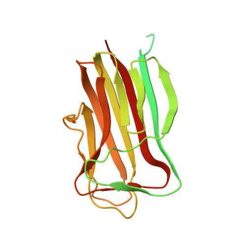
    The lethal disease anthrax is propagated by spores of Bacillus anthracis, which can penetrate into the mammalian host by inhalation, causing a rapid progression of the disease and a mostly fatal outcome. We have solved the three-dimensional structure of the major surface protein BclA on B. anthracis spores. Surprisingly, the structure resembles C1q, the first component of complement, despite there being no sequence homology. Although most assays for C1q-like activity, including binding to C1q receptors, suggest that BclA does not mimic C1q, we show that BclA, as well as C1q, interacts with components of the lung alveolar surfactant layer. Thus, to better recognize and invade its hosts, this pathogenic soil bacterium may have evolved a surface protein whose structure is strikingly close to a mammalian protein.


  • Organizational Affiliation
    • Laboratoire de Biotechnologies et Pharmacologie Génétique Appliquées, CNRS, Unité Mixte de Recherche 8113, Ecole Normale Supérieure de Cachan, 61 Avenue du Président Wilson, 94235 Cachan, France.

Macromolecule Content 

  • Total Structure Weight: 21.53 kDa 
  • Atom Count: 1,170 
  • Modeled Residue Count: 136 
  • Deposited Residue Count: 220 
  • Unique protein chains: 1

Macromolecules

Find similar proteins by:|  3D Structure
Entity ID: 1
MoleculeChains  Sequence LengthOrganismDetailsImage
BCLA PROTEIN220Bacillus anthracisMutation(s): 0 
UniProt
Find proteins for Q83WB0 (Bacillus anthracis)
Explore Q83WB0 
Go to UniProtKB:  Q83WB0
Entity Groups
Sequence Clusters30% Identity50% Identity70% Identity90% Identity95% Identity100% Identity
UniProt GroupQ83WB0
Sequence Annotations
Expand
Reference Sequence

Small Molecules

Ligands 1 Unique
IDChains Name / Formula / InChI Key2D Diagram3D Interactions
CAC

Query on CAC


Download:Ideal Coordinates CCD File
B [auth A]CACODYLATE ION
C2 H6 As O2
OGGXGZAMXPVRFZ-UHFFFAOYSA-M

Experimental Data & Validation

Experimental Data

  • Method: X-RAY DIFFRACTION
  • Resolution: 1.36 Å
  • R-Value Free:  0.190 (Depositor), 0.188 (DCC) 
  • R-Value Work:  0.179 (Depositor) 
  • R-Value Observed: 0.179 (Depositor) 
Space Group: P 63 2 2
Unit Cell:
Length ( Å )Angle ( ˚ )
a = 68.558α = 90
b = 68.558β = 90
c = 163.35γ = 120
Software Package:
Software NamePurpose
REFMACrefinement
XDSdata reduction
XSCALEdata scaling
autoSHARPphasing

Structure Validation

View Full Validation Report


Entry History 

Deposition Data

Revision History  (Full details and data files)

  • Version 1.0: 2005-10-25
    Type: Initial release
  • Version 1.1: 2011-05-08
    Changes: Version format compliance
  • Version 1.2: 2011-07-13
    Changes: Version format compliance
  • Version 1.3: 2019-03-06
    Changes: Data collection, Derived calculations, Experimental preparation, Other
  • Version 1.4: 2024-10-16
    Changes: Data collection, Database references, Other, Structure summary