1W8A | pdb_00001w8a

Third LRR domain of Drosophila Slit


Experimental Data Snapshot

  • Method: X-RAY DIFFRACTION
  • Resolution: 2.80 Å
  • R-Value Free: 
    0.269 (Depositor), 0.267 (DCC) 
  • R-Value Work: 
    0.199 (Depositor), 0.199 (DCC) 
  • R-Value Observed: 
    0.199 (Depositor) 

Starting Model: experimental
View more details

wwPDB Validation 3D Report Full Report

Validation slider image for 1W8A

This is version 1.5 of the entry. See complete history

Literature

Binding Site for Robo Receptors Revealed by Dissection of the Leucine-Rich Repeat Region of Slit.

Howitt, J.A.Clout, N.J.Hohenester, E.

(2004) EMBO J 23: 4406

  • DOI: https://doi.org/10.1038/sj.emboj.7600446
  • Primary Citation Related Structures: 
    1W8A

  • PubMed Abstract: 

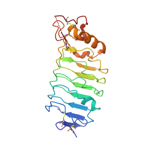
    Recognition of the large secreted protein Slit by receptors of the Robo family provides fundamental signals in axon guidance and other developmental processes. In Drosophila, Slit-Robo signalling regulates midline crossing and the lateral position of longitudinal axon tracts. We report the functional dissection of Drosophila Slit, using structure analysis, site-directed mutagenesis and in vitro assays. The N-terminal region of Slit consists of a tandem array of four independently folded leucine-rich repeat (LRR) domains, connected by disulphide-tethered linkers. All three Drosophila Robos were found to compete for a single highly conserved site on the concave face of the second LRR domain of Slit. We also found that this domain is sufficient for biological activity in a chemotaxis assay. Other Slit activities may require Slit dimerisation mediated by the fourth LRR domain. Our results show that a small portion of Slit is able to induce Robo signalling and indicate that the distinct functions of Drosophila Robos are encoded in their divergent cytosolic domains.


  • Organizational Affiliation
    • Department of Biological Sciences, Imperial College London, London, UK. e.hohenester@imperial.ac.uk

Macromolecule Content 

  • Total Structure Weight: 21.31 kDa 
  • Atom Count: 1,470 
  • Modeled Residue Count: 189 
  • Deposited Residue Count: 192 
  • Unique protein chains: 1

Macromolecules

Find similar proteins by:|  3D Structure
Entity ID: 1
MoleculeChains  Sequence LengthOrganismDetailsImage
SLIT PROTEIN192Drosophila melanogasterMutation(s): 0 
UniProt
Find proteins for P24014 (Drosophila melanogaster)
Explore P24014 
Go to UniProtKB:  P24014
Entity Groups
Sequence Clusters30% Identity50% Identity70% Identity90% Identity95% Identity100% Identity
UniProt GroupP24014
Sequence Annotations
Expand
Reference Sequence

Experimental Data & Validation

Experimental Data

  • Method: X-RAY DIFFRACTION
  • Resolution: 2.80 Å
  • R-Value Free:  0.269 (Depositor), 0.267 (DCC) 
  • R-Value Work:  0.199 (Depositor), 0.199 (DCC) 
  • R-Value Observed: 0.199 (Depositor) 
Space Group: C 1 2 1
Unit Cell:
Length ( Å )Angle ( ˚ )
a = 121.213α = 90
b = 31.852β = 100.72
c = 49.526γ = 90
Software Package:
Software NamePurpose
CNSrefinement
MOSFLMdata reduction
SCALAdata scaling
AMoREphasing

Structure Validation

View Full Validation Report


Entry History 

Deposition Data

Revision History  (Full details and data files)

  • Version 1.0: 2004-10-25
    Type: Initial release
  • Version 1.1: 2011-05-08
    Changes: Version format compliance
  • Version 1.2: 2011-07-13
    Changes: Version format compliance
  • Version 1.3: 2017-11-08
    Changes: Advisory, Source and taxonomy
  • Version 1.4: 2023-12-13
    Changes: Advisory, Data collection, Database references, Other, Refinement description
  • Version 1.5: 2024-11-20
    Changes: Structure summary