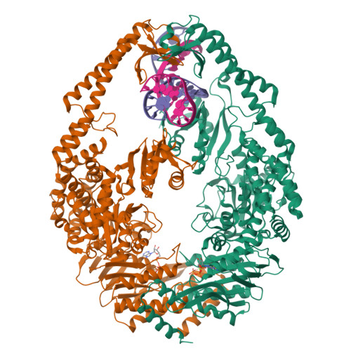
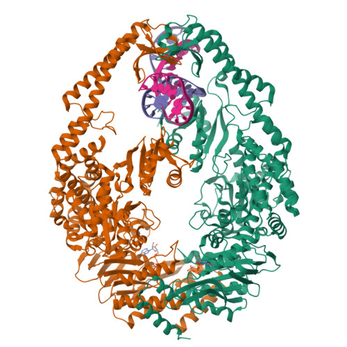
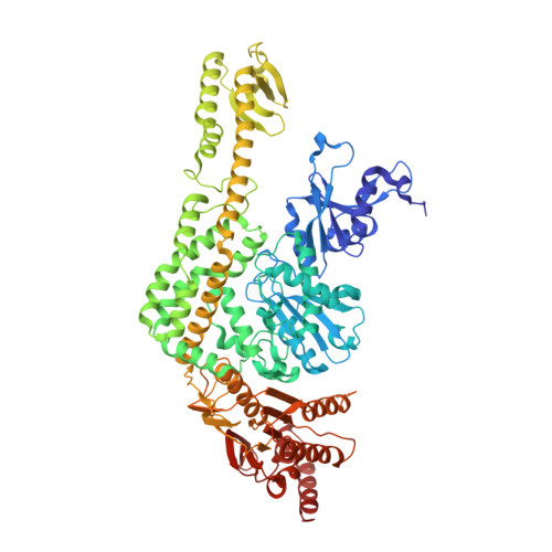
1W7A

ATP bound MutS


Experimental Data Snapshot

  • Method: X-RAY DIFFRACTION
  • Resolution: 2.27 Å
  • R-Value Free: 0.253 
  • R-Value Work: 0.216 
  • R-Value Observed: 0.217 

Starting Model: experimental
View more details

wwPDB Validation   3D Report Full Report


Ligand Structure Quality Assessment 


This is version 1.2 of the entry. See complete history


Literature

ATP Increases the Affinity between Muts ATPase Domains: Implications for ATP Hydrolysis and Conformational Changes

Lamers, M.H.Georgijevic, D.Lebbink, J.Winterwerp, H.H.K.Agianian, B.De Wind, N.Sixma, T.K.

(2004) J Biol Chem 279: 43879

  • DOI: https://doi.org/10.1074/jbc.M406380200
  • Primary Citation of Related Structures:  
    1W7A

  • PubMed Abstract: 

    MutS is the key protein of the Escherichia coli DNA mismatch repair system. It recognizes mispaired and unpaired bases and has intrinsic ATPase activity. ATP binding after mismatch recognition by MutS serves as a switch that enables MutL binding and the subsequent initiation of mismatch repair. However, the mechanism of this switch is poorly understood. We have investigated the effects of ATP binding on the MutS structure. Crystallographic studies of ATP-soaked crystals of MutS show a trapped intermediate, with ATP in the nucleotide-binding site. Local rearrangements of several residues around the nucleotide-binding site suggest a movement of the two ATPase domains of the MutS dimer toward each other. Analytical ultracentrifugation experiments confirm such a rearrangement, showing increased affinity between the ATPase domains upon ATP binding and decreased affinity in the presence of ADP. Mutations of specific residues in the nucleotide-binding domain reduce the dimer affinity of the ATPase domains. In addition, ATP-induced release of DNA is strongly reduced in these mutants, suggesting that the two activities are coupled. Hence, it seems plausible that modulation of the affinity between ATPase domains is the driving force for conformational changes in the MutS dimer. These changes are driven by distinct amino acids in the nucleotide-binding site and form the basis for long-range interactions between the ATPase domains and DNA-binding domains and subsequent binding of MutL and initiation of mismatch repair.


  • Organizational Affiliation

    Division of Molecular Carcinogenesis, Netherlands Cancer Institute, Plesmanlaan 121, 1066 CX Amsterdam.


Macromolecules

Find similar proteins by:  (by identity cutoff)  |  3D Structure
Entity ID: 1
MoleculeChains Sequence LengthOrganismDetailsImage
DNA MISMATCH REPAIR PROTEIN MUTS
A, B
800Escherichia coliMutation(s): 0 
UniProt
Find proteins for P23909 (Escherichia coli (strain K12))
Explore P23909 
Go to UniProtKB:  P23909
Entity Groups  
Sequence Clusters30% Identity50% Identity70% Identity90% Identity95% Identity100% Identity
UniProt GroupP23909
Sequence Annotations
Expand
  • Reference Sequence
Find similar nucleic acids by:  (by identity cutoff)  |  3D Structure
Entity ID: 2
MoleculeChains LengthOrganismImage
5'-D(*AP*GP*CP*TP*GP*CP*CP*AP*GP*GP *CP*AP*CP*CP*AP*GP*TP*GP*TP*CP*AP*GP*CP*GP*TP*CP*CP*TP* AP*T)-3'C [auth E]30N/A
Sequence Annotations
Expand
  • Reference Sequence
Find similar nucleic acids by:  (by identity cutoff)  |  3D Structure
Entity ID: 3
MoleculeChains LengthOrganismImage
5'-D(*AP*TP*AP*GP*GP*AP*CP*GP*CP*TP *GP*AP*CP*AP*CP*TP*GP*GP*TP*GP*CP*TP*TP*GP*GP*CP*AP*GP* CP*T)-3'D [auth F]30N/A
Sequence Annotations
Expand
  • Reference Sequence
Experimental Data & Validation

Experimental Data

  • Method: X-RAY DIFFRACTION
  • Resolution: 2.27 Å
  • R-Value Free: 0.253 
  • R-Value Work: 0.216 
  • R-Value Observed: 0.217 
  • Space Group: P 21 21 21
Unit Cell:
Length ( Å )Angle ( ˚ )
a = 90.636α = 90
b = 93.04β = 90
c = 262.592γ = 90
Software Package:
Software NamePurpose
REFMACrefinement
HKL-2000data reduction
HKL-2000data scaling
AMoREphasing

Structure Validation

View Full Validation Report


Ligand Structure Quality Assessment 


Entry History 

Deposition Data

Revision History  (Full details and data files)

  • Version 1.0: 2004-09-10
    Type: Initial release
  • Version 1.1: 2011-07-13
    Changes: Advisory, Version format compliance
  • Version 1.2: 2023-12-13
    Changes: Data collection, Database references, Derived calculations, Other, Refinement description