1W5Z | pdb_00001w5z

AGAO covalent complex with Benzylhydrazine


Experimental Data Snapshot

  • Method: X-RAY DIFFRACTION
  • Resolution: 1.86 Å
  • R-Value Free: 
    0.187 (Depositor), 0.192 (DCC) 
  • R-Value Work: 
    0.152 (Depositor), 0.159 (DCC) 
  • R-Value Observed: 
    0.154 (Depositor) 

Starting Model: experimental
View more details

wwPDB Validation 3D Report Full Report

Validation slider image for 1W5Z

This is version 3.0 of the entry. See complete history

Literature

Complexes of the Copper-Containing Amine Oxidase from Arthrobacter Globiformis with the Inhibitors Benzylhydrazine and Tranylcypromine.

Langley, D.B.Trambaiolo, D.M.Duff, A.P.Dooley, D.M.Freeman, H.C.Guss, J.M.

(2008) Acta Crystallogr Sect F Struct Biol Cryst Commun 64: 577

  • DOI: https://doi.org/10.1107/S174430910801556X
  • Primary Citation Related Structures: 
    1W4N, 1W5Z

  • PubMed Abstract: 

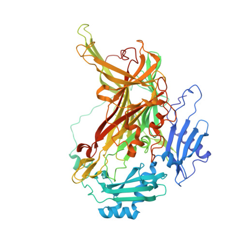
    Complexes of Arthrobacter globiformis amine oxidase (AGAO) with the inhibitors benzylhydrazine and tranylcypromine (an antidepressant drug) have been refined at 1.86 and 1.65 A resolution, respectively. Both inhibitors form covalent adducts with the TPQ cofactor. A tyrosine residue, proposed to act as a gate to the AGAO active site, is in its open conformation.


  • Organizational Affiliation
    • School of Molecular and Microbial Biosciences, University of Sydney, Australia.

Macromolecule Content 

  • Total Structure Weight: 72.7 kDa 
  • Atom Count: 5,431 
  • Modeled Residue Count: 621 
  • Deposited Residue Count: 646 
  • Unique protein chains: 1

Macromolecules

Find similar proteins by:|  3D Structure
Entity ID: 1
MoleculeChains  Sequence LengthOrganismDetailsImage
PHENYLETHYLAMINE OXIDASE646Arthrobacter globiformisMutation(s): 0 
EC: 1.4.3.6 (PDB Primary Data), 1.4.3.21 (UniProt)
UniProt
Find proteins for P46881 (Arthrobacter globiformis)
Explore P46881 
Go to UniProtKB:  P46881
Entity Groups
Sequence Clusters30% Identity50% Identity70% Identity90% Identity95% Identity100% Identity
UniProt GroupP46881
Sequence Annotations
Expand
Reference Sequence

Small Molecules

Ligands 4 Unique
IDChains Name / Formula / InChI Key2D Diagram3D Interactions
SO4

Query on SO4


Download:Ideal Coordinates CCD File
D [auth A],
E [auth A],
F [auth A]
SULFATE ION
O4 S
QAOWNCQODCNURD-UHFFFAOYSA-L
GOL

Query on GOL


Download:Ideal Coordinates CCD File
G [auth A],
H [auth A],
I [auth A],
J [auth A],
K [auth A]
GLYCEROL
C3 H8 O3
PEDCQBHIVMGVHV-UHFFFAOYSA-N
CU

Query on CU


Download:Ideal Coordinates CCD File
B [auth A]COPPER (II) ION
Cu
JPVYNHNXODAKFH-UHFFFAOYSA-N
NA

Query on NA


Download:Ideal Coordinates CCD File
C [auth A]SODIUM ION
Na
FKNQFGJONOIPTF-UHFFFAOYSA-N
Modified Residues  1 Unique
IDChains TypeFormula2D DiagramParent
3TY
Query on 3TY
A
L-PEPTIDE LINKINGC16 H17 N3 O4TYR

Experimental Data & Validation

Experimental Data

  • Method: X-RAY DIFFRACTION
  • Resolution: 1.86 Å
  • R-Value Free:  0.187 (Depositor), 0.192 (DCC) 
  • R-Value Work:  0.152 (Depositor), 0.159 (DCC) 
  • R-Value Observed: 0.154 (Depositor) 
Space Group: C 1 2 1
Unit Cell:
Length ( Å )Angle ( ˚ )
a = 158.126α = 90
b = 62.665β = 112.07
c = 92.283γ = 90
Software Package:
Software NamePurpose
REFMACrefinement
DENZOdata reduction
SCALEPACKdata scaling

Structure Validation

View Full Validation Report


Entry History 

Deposition Data

Revision History  (Full details and data files)

  • Version 1.0: 2005-12-08
    Type: Initial release
  • Version 1.1: 2011-07-13
    Changes: Advisory, Version format compliance
  • Version 1.2: 2013-08-07
    Changes: Non-polymer description, Other, Source and taxonomy
  • Version 1.3: 2019-03-06
    Changes: Data collection, Derived calculations, Experimental preparation, Other
  • Version 1.4: 2019-05-15
    Changes: Data collection, Experimental preparation
  • Version 2.0: 2023-11-15
    Changes: Atomic model, Data collection, Database references, Derived calculations, Other
  • Version 2.1: 2023-12-13
    Changes: Refinement description
  • Version 3.0: 2025-07-30
    Changes: Polymer sequence, Structure summary