1W4S | pdb_00001w4s

Crystal structure of the proximal BAH domain of polybromo


Experimental Data Snapshot

  • Method: X-RAY DIFFRACTION
  • Resolution: 1.55 Å
  • R-Value Free: 
    0.230 (Depositor), 0.232 (DCC) 
  • R-Value Work: 
    0.199 (Depositor), 0.201 (DCC) 
  • R-Value Observed: 
    0.201 (Depositor) 

wwPDB Validation 3D Report Full Report

Validation slider image for 1W4S

This is version 1.2 of the entry. See complete history

Literature

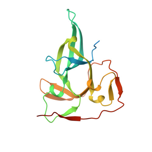
Crystal Structure of the Proximal Bah Domain of the Polybromo Protein

Oliver, A.W.Jones, S.A.Roe, S.M.Matthews, S.Goodwin, G.H.Pearl, L.H.

(2005) Biochem J 389: 657

  • DOI: https://doi.org/10.1042/BJ20050310
  • Primary Citation Related Structures: 
    1W4S

  • PubMed Abstract: 

    The BAH domain (bromo-associated homology domain) was first identified from a repeated motif found in the nuclear protein polybromo--a large (187 kDa) modular protein comprising six bromodomains, two BAH domains and an HMG box. To date, the BAH domain has no ascribed function, although it is found in a wide range of proteins that contain additional domains involved in either transcriptional regulation (e.g. SET, PHD and bromodomain) and/or DNA binding (HMG box and AT hook). The molecular function of polybromo itself also remains unclear, but it has been identified as a key component of an SWI/SNF (switching/sucrose non-fermenting)-related, ATP-dependent chromatin-remodelling complex PBAF (polybromo, BRG1-associated factors; also known as SWI/SNF-B or SWI/SNFbeta). We present in this paper the crystal structure of the proximal BAH domain from chicken polybromo (BAH1), at a resolution of 1.6 A (1 A=0.1 nm). Structure-based sequence analysis reveals several features that may be involved in mediating protein-protein interactions.


  • Organizational Affiliation
    • CR-UK DNA Repair Enzymes Group, Section of Structural Biology, The Institute of Cancer Research, 237 Fulham Road, Chelsea, London SW3 6JB, UK. antony.oliver@icr.ac.uk

Macromolecule Content 

  • Total Structure Weight: 20.36 kDa 
  • Atom Count: 1,327 
  • Modeled Residue Count: 146 
  • Deposited Residue Count: 174 
  • Unique protein chains: 1

Macromolecules

Find similar proteins by:|  3D Structure
Entity ID: 1
MoleculeChains  Sequence LengthOrganismDetailsImage
POLYBROMO 1 PROTEIN174Gallus gallusMutation(s): 0 
UniProt
Find proteins for Q90941 (Gallus gallus)
Explore Q90941 
Go to UniProtKB:  Q90941
Entity Groups
Sequence Clusters30% Identity50% Identity70% Identity90% Identity95% Identity100% Identity
UniProt GroupQ90941
Sequence Annotations
Expand
Reference Sequence

Small Molecules

Ligands 1 Unique
IDChains Name / Formula / InChI Key2D Diagram3D Interactions
CL

Query on CL


Download:Ideal Coordinates CCD File
B [auth A]CHLORIDE ION
Cl
VEXZGXHMUGYJMC-UHFFFAOYSA-M

Experimental Data & Validation

Experimental Data

  • Method: X-RAY DIFFRACTION
  • Resolution: 1.55 Å
  • R-Value Free:  0.230 (Depositor), 0.232 (DCC) 
  • R-Value Work:  0.199 (Depositor), 0.201 (DCC) 
  • R-Value Observed: 0.201 (Depositor) 
Space Group: P 21 21 21
Unit Cell:
Length ( Å )Angle ( ˚ )
a = 39.598α = 90
b = 57.712β = 90
c = 70.377γ = 90
Software Package:
Software NamePurpose
REFMACrefinement
MOSFLMdata reduction
SCALAdata scaling
SHARPphasing

Structure Validation

View Full Validation Report


Entry History 

Deposition Data

Revision History  (Full details and data files)

  • Version 1.0: 2005-04-27
    Type: Initial release
  • Version 1.1: 2011-07-13
    Changes: Advisory, Version format compliance
  • Version 1.2: 2024-05-08
    Changes: Data collection, Database references, Other