1W30 | pdb_00001w30

PyrR of Mycobacterium Tuberculosis as a potential drug target


Experimental Data Snapshot

  • Method: X-RAY DIFFRACTION
  • Resolution: 1.90 Å
  • R-Value Free: 
    0.244 (Depositor), 0.252 (DCC) 
  • R-Value Work: 
    0.205 (Depositor), 0.216 (DCC) 
  • R-Value Observed: 
    0.206 (Depositor) 

Starting Model: experimental
View more details

wwPDB Validation 3D Report Full Report

Validation slider image for 1W30

This is version 1.4 of the entry. See complete history

Literature

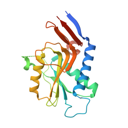
Structure of Pyrr (Rv1379) from Mycobacterium Tuberculosis: A Persistence Gene and Protein Drug Target

Kantardjieff, K.A.Vasquez, C.Castro, P.Warfel, N.N.Rho, B.-S.Lekin, T.Kim, C.-Y.Segelke, B.W.Terwilliger, T.Rupp, B.

(2005) Acta Crystallogr D Biol Crystallogr 61: 355

  • DOI: https://doi.org/10.1107/S090744490403389X
  • Primary Citation Related Structures: 
    1W30

  • PubMed Abstract: 

    The Mycobacterium tuberculosis pyrR gene (Rv1379) encodes a protein that regulates the expression of pyrimidine-nucleotide biosynthesis (pyr) genes in a UMP-dependent manner. Because pyrimidine biosynthesis is an essential step in the progression of TB, the gene product pyrR is an attractive antitubercular drug target. The 1.9 A native structure of Mtb pyrR determined by the TB Structural Genomics Consortium facilities in trigonal space group P3(1)21 is reported, with unit-cell parameters a = 66.64, c = 154.72 A at 120 K and two molecules in the asymmetric unit. The three-dimensional structure and residual uracil phosphoribosyltransferase activity point to a common PRTase ancestor for pyrR. However, while PRPP- and UMP-binding sites have been retained in Mtb pyrR, a distinct dimer interaction among subunits creates a deep positively charged cleft capable of binding pyr mRNA. In silico screening of pyrimidine-nucleoside analogs has revealed a number of potential lead compounds that, if bound to Mtb pyrR, could facilitate transcriptional attenuation, particularly cyclopentenyl nucleosides.


  • Organizational Affiliation
    • Department of Chemistry and Biochemistry and W. M. Keck Foundation Center for Molecular Structure, California State University Fullerton, Fullerton, CA 92834, USA.

Macromolecule Content 

  • Total Structure Weight: 43.26 kDa 
  • Atom Count: 2,985 
  • Modeled Residue Count: 351 
  • Deposited Residue Count: 402 
  • Unique protein chains: 1

Macromolecules

Find similar proteins by:|  3D Structure
Entity ID: 1
MoleculeChains  Sequence LengthOrganismDetailsImage
PYRR BIFUNCTIONAL PROTEIN
A, B
201Mycobacterium tuberculosisMutation(s): 1 
EC: 2.4.2.9
UniProt
Find proteins for P9WHK3 (Mycobacterium tuberculosis (strain ATCC 25618 / H37Rv))
Explore P9WHK3 
Go to UniProtKB:  P9WHK3
Entity Groups
Sequence Clusters30% Identity50% Identity70% Identity90% Identity95% Identity100% Identity
UniProt GroupP9WHK3
Sequence Annotations
Expand
Reference Sequence

Experimental Data & Validation

Experimental Data

  • Method: X-RAY DIFFRACTION
  • Resolution: 1.90 Å
  • R-Value Free:  0.244 (Depositor), 0.252 (DCC) 
  • R-Value Work:  0.205 (Depositor), 0.216 (DCC) 
  • R-Value Observed: 0.206 (Depositor) 
Space Group: P 31 2 1
Unit Cell:
Length ( Å )Angle ( ˚ )
a = 66.638α = 90
b = 66.638β = 90
c = 154.716γ = 120
Software Package:
Software NamePurpose
REFMACrefinement
DENZOdata reduction
SCALEPACKdata scaling
EPMRphasing

Structure Validation

View Full Validation Report


Entry History 

Revision History  (Full details and data files)

  • Version 1.0: 2004-09-29
    Type: Initial release
  • Version 1.1: 2015-05-06
    Changes: Derived calculations, Non-polymer description, Other, Structure summary, Version format compliance
  • Version 1.2: 2019-05-08
    Changes: Data collection, Experimental preparation, Other
  • Version 1.3: 2019-08-21
    Changes: Data collection, Database references
  • Version 1.4: 2023-12-13
    Changes: Data collection, Database references, Other, Refinement description