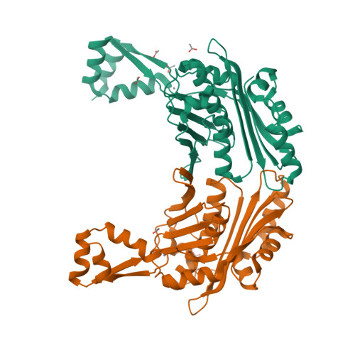
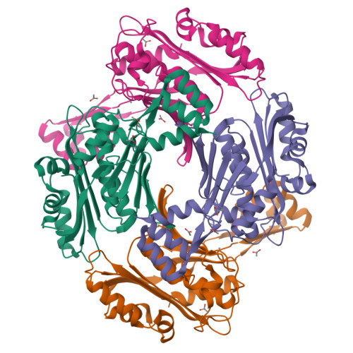
The crystal structure of the reduced, Zn2+-bound form of the B. subtilis Hsp33 chaperone and its implications for the activation mechanism.
Janda, I., Devedjiev, Y., Derewenda, U., Dauter, Z., Bielnicki, J., Cooper, D.R., Graf, P.C., Joachimiak, A., Jakob, U., Derewenda, Z.S.(2004) Structure 12: 1901-1907
- PubMed: 15458638
- DOI: https://doi.org/10.1016/j.str.2004.08.003
- Primary Citation of Related Structures:
1VZY - PubMed Abstract:
The bacterial heat shock protein Hsp33 is a redox-regulated chaperone activated by oxidative stress. In response to oxidation, four cysteines within a Zn2+ binding C-terminal domain form two disulfide bonds with concomitant release of the metal. This leads to the formation of the biologically active Hsp33 dimer. The crystal structure of the N-terminal domain of the E. coli protein has been reported, but neither the structure of the Zn2+ binding motif nor the nature of its regulatory interaction with the rest of the protein are known. Here we report the crystal structure of the full-length B. subtilis Hsp33 in the reduced form. The structure of the N-terminal, dimerization domain is similar to that of the E. coli protein, although there is no domain swapping. The Zn2+ binding domain is clearly resolved showing the details of the tetrahedral coordination of Zn2+ by four thiolates. We propose a structure-based activation pathway for Hsp33.
Organizational Affiliation:
Department of Molecular Physiology and Biological Physics, University of Virginia, Charlottesville, VA 22908, USA.


