1VT6

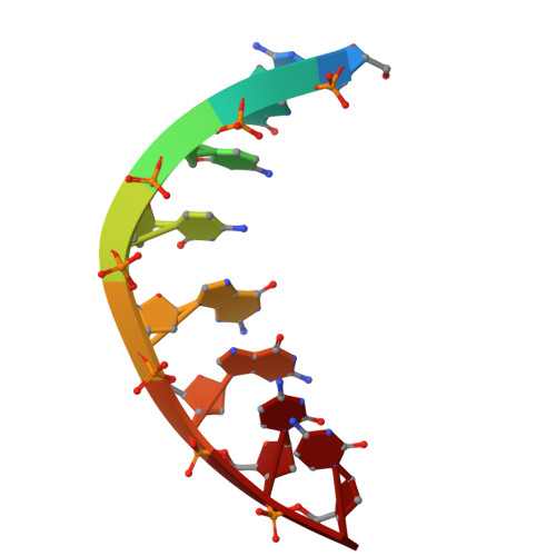
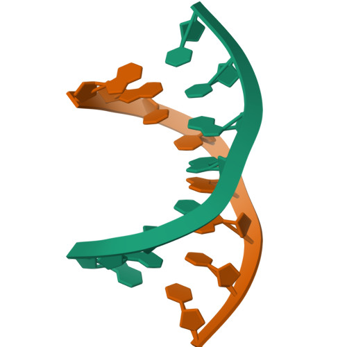
Molecular structure of the octamer D(G-G-C-C-G-G-C-C) modified A-DNA


Experimental Data Snapshot

  • Method: X-RAY DIFFRACTION
  • Resolution: 2.25 Å
  • R-Value Work: 0.174 
  • R-Value Observed: 0.174 

wwPDB Validation   3D Report Full Report


This is version 1.1 of the entry. See complete history


Literature

Molecular structure of the octamer d(G-G-C-C-G-G-C-C): modified A-DNA.

Wang, A.H.Fujii, S.van Boom, J.H.Rich, A.

(1982) Proc Natl Acad Sci U S A 79: 3968-3972

  • DOI: https://doi.org/10.1073/pnas.79.13.3968
  • Primary Citation of Related Structures:  
    1VT6

  • PubMed Abstract: 

    The deoxynucleotide fragment d(GpGpCpCpGpGpCpC) was synthesized and crystallized, and its three-dimensional structure was determined by x-ray diffraction techniques to a resolution of 2.25 A. The molecule forms a right-handed double helix in which the two base pairs at either end of the molecule are in the conventional A-DNA conformation, while the central four base pairs have a modified form in which alternate residues have sugar conformations that are closer to those in B-DNA than in A-DNA. The molecules have an intermolecular contact in which the planar terminal guanine . cytosine base pair lies on the flat minor groove surface of the A-DNA helix.


Macromolecules

Find similar nucleic acids by:  Sequence   |   3D Structure  

Entity ID: 1
MoleculeChains LengthOrganismImage
DNA (5'-D(*GP*GP*CP*CP*GP*GP*CP*C)-3')8N/A
Sequence Annotations
Expand
  • Reference Sequence
Experimental Data & Validation

Experimental Data

  • Method: X-RAY DIFFRACTION
  • Resolution: 2.25 Å
  • R-Value Work: 0.174 
  • R-Value Observed: 0.174 
  • Space Group: P 43 21 2
Unit Cell:
Length ( Å )Angle ( ˚ )
a = 42.06α = 90
b = 42.06β = 90
c = 25.17γ = 90

Structure Validation

View Full Validation Report


Entry History 

Deposition Data

Revision History  (Full details and data files)

  • Version 1.0: 2011-07-13
    Type: Initial release
  • Version 1.1: 2023-12-27
    Changes: Data collection, Database references