1VP6 | pdb_00001vp6

M.loti ion channel cylic nucleotide binding domain


Experimental Data Snapshot

  • Method: X-RAY DIFFRACTION
  • Resolution: 1.70 Å
  • R-Value Free: 
    0.219 (Depositor), 0.234 (DCC) 
  • R-Value Work: 
    0.190 (Depositor), 0.205 (DCC) 
  • R-Value Observed: 
    0.219 (Depositor) 

wwPDB Validation 3D Report Full Report

Validation slider image for 1VP6

Ligand Structure Quality Assessment 


This is version 1.3 of the entry. See complete history

Literature

Structural basis of ligand activation in a cyclic nucleotide regulated potassium channel.

Clayton, G.M.Silverman, W.R.Heginbotham, L.Morais-Cabral, J.H.

(2004) Cell 119: 615-627

  • DOI: https://doi.org/10.1016/j.cell.2004.10.030
  • Primary Citation Related Structures: 
    1U12, 1VP6

  • PubMed Abstract: 

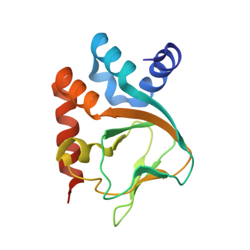
    Here we describe the initial functional characterization of a cyclic nucleotide regulated ion channel from the bacterium Mesorhizobium loti and present two structures of its cyclic nucleotide binding domain, with and without cAMP. The domains are organized as dimers with the interface formed by the linker regions that connect the nucleotide binding pocket to the pore domain. Together, structural and functional data suggest the domains form two dimers on the cytoplasmic face of the channel. We propose a model for gating in which ligand binding alters the structural relationship within a dimer, directly affecting the position of the adjacent transmembrane helices.


  • Organizational Affiliation
    • Department of Molecular Biophysics and Biochemistry, Yale University, 260 Whitney Avenue, New Haven, CT 06520, USA.

Macromolecule Content 

  • Total Structure Weight: 31.1 kDa 
  • Atom Count: 2,316 
  • Modeled Residue Count: 266 
  • Deposited Residue Count: 276 
  • Unique protein chains: 1

Macromolecules

Find similar proteins by:|  3D Structure
Entity ID: 1
MoleculeChains  Sequence LengthOrganismDetailsImage
Cyclic-nucleotide binding domain of mesorhizobium loti CNG potassium channelA,
B [auth C]
138Mesorhizobium lotiMutation(s): 0 
UniProt
Find proteins for Q98GN8 (Mesorhizobium japonicum (strain LMG 29417 / CECT 9101 / MAFF 303099))
Explore Q98GN8 
Go to UniProtKB:  Q98GN8
Entity Groups
Sequence Clusters30% Identity50% Identity70% Identity90% Identity95% Identity100% Identity
UniProt GroupQ98GN8
Sequence Annotations
Expand
Reference Sequence

Small Molecules

Ligands 2 Unique
IDChains Name / Formula / InChI Key2D Diagram3D Interactions
CMP

Query on CMP


Download:Ideal Coordinates CCD File
O [auth A],
T [auth C]
ADENOSINE-3',5'-CYCLIC-MONOPHOSPHATE
C10 H12 N5 O6 P
IVOMOUWHDPKRLL-KQYNXXCUSA-N
BR

Query on BR


Download:Ideal Coordinates CCD File
C [auth A]
D [auth A]
E [auth A]
F [auth A]
G [auth A]
C [auth A],
D [auth A],
E [auth A],
F [auth A],
G [auth A],
H [auth A],
I [auth A],
J [auth A],
K [auth A],
L [auth A],
M [auth A],
N [auth A],
P [auth C],
Q [auth C],
R [auth C],
S [auth C]
BROMIDE ION
Br
CPELXLSAUQHCOX-UHFFFAOYSA-M

Experimental Data & Validation

Experimental Data

  • Method: X-RAY DIFFRACTION
  • Resolution: 1.70 Å
  • R-Value Free:  0.219 (Depositor), 0.234 (DCC) 
  • R-Value Work:  0.190 (Depositor), 0.205 (DCC) 
  • R-Value Observed: 0.219 (Depositor) 
Space Group: P 1 21 1
Unit Cell:
Length ( Å )Angle ( ˚ )
a = 34.324α = 90
b = 80.292β = 99.16
c = 50.419γ = 90
Software Package:
Software NamePurpose
CNSrefinement
MOSFLMdata reduction
CCP4data scaling
SOLVEphasing
RESOLVEphasing

Structure Validation

View Full Validation Report


Ligand Structure Quality Assessment 


Entry History 

Deposition Data

Revision History  (Full details and data files)

  • Version 1.0: 2004-10-26
    Type: Initial release
  • Version 1.1: 2008-04-26
    Changes: Version format compliance
  • Version 1.2: 2011-07-13
    Changes: Version format compliance
  • Version 1.3: 2023-12-27
    Changes: Data collection, Database references, Derived calculations