1VOT | pdb_00001vot

ACETYLCHOLINESTERASE (E.C. 3.1.1.7) COMPLEXED WITH HUPERZINE A


Experimental Data Snapshot

  • Method: X-RAY DIFFRACTION
  • Resolution: 2.50 Å
  • R-Value Free: 
    0.248 (Depositor), 0.220 (DCC) 
  • R-Value Work: 
    0.205 (Depositor), 0.182 (DCC) 
  • R-Value Observed: 
    0.205 (Depositor) 

Starting Model: experimental
View more details

wwPDB Validation 3D Report Full Report

Validation slider image for 1VOT

Ligand Structure Quality Assessment 


This is version 1.5 of the entry. See complete history

Literature

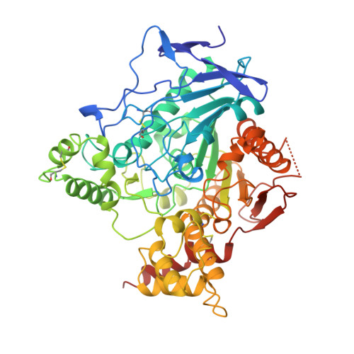
Structure of acetylcholinesterase complexed with the nootropic alkaloid, (-)-huperzine A.

Raves, M.L.Harel, M.Pang, Y.P.Silman, I.Kozikowski, A.P.Sussman, J.L.

(1997) Nat Struct Biol 4: 57-63

  • DOI: https://doi.org/10.1038/nsb0197-57
  • Primary Citation Related Structures: 
    1VOT, 2ACE

  • PubMed Abstract: 

    (-)-Huperzine A (HupA) is found in an extract from a club moss that has been used for centuries in Chinese folk medicine. Its action has been attributed to its ability to strongly inhibit acetylcholinesterase (AChE). The crystal structure of the complex of AChE with optically pure HupA at 2.5 A resolution shows an unexpected orientation for the inhibitor with surprisingly few strong direct interactions with protein residues to explain its high affinity. This structure is compared to the native structure of AChE devoid of any inhibitor as determined to the same resolution. An analysis of the affinities of structural analogues of HupA, correlated with their interactions with the protein, shows the importance of individual hydrophobic interactions between HupA and aromatic residues in the active-site gorge of AChE.


  • Organizational Affiliation
    • Department of Structural Biology, Weizmann Institute of Science, Rehovot, Israel.

Macromolecule Content 

  • Total Structure Weight: 60.98 kDa 
  • Atom Count: 4,366 
  • Modeled Residue Count: 529 
  • Deposited Residue Count: 537 
  • Unique protein chains: 1

Macromolecules

Find similar proteins by:|  3D Structure
Entity ID: 1
MoleculeChains  Sequence LengthOrganismDetailsImage
ACETYLCHOLINESTERASE537Tetronarce californicaMutation(s): 0 
EC: 3.1.1.7
UniProt
Find proteins for P04058 (Tetronarce californica)
Explore P04058 
Go to UniProtKB:  P04058
Entity Groups
Sequence Clusters30% Identity50% Identity70% Identity90% Identity95% Identity100% Identity
UniProt GroupP04058
Sequence Annotations
Expand
Reference Sequence

Small Molecules

Ligands 1 Unique
IDChains Name / Formula / InChI Key2D Diagram3D Interactions
HUP

Query on HUP


Download:Ideal Coordinates CCD File
B [auth A]Huperzine A
C15 H18 N2 O
ZRJBHWIHUMBLCN-YQEJDHNASA-N
Binding Affinity Annotations 
IDSourceBinding Affinity
HUP BindingDB:  1VOT IC50: 11.4 (nM) from 1 assay(s)

Experimental Data & Validation

Experimental Data

  • Method: X-RAY DIFFRACTION
  • Resolution: 2.50 Å
  • R-Value Free:  0.248 (Depositor), 0.220 (DCC) 
  • R-Value Work:  0.205 (Depositor), 0.182 (DCC) 
  • R-Value Observed: 0.205 (Depositor) 
Space Group: P 31 2 1
Unit Cell:
Length ( Å )Angle ( ˚ )
a = 112.632α = 90
b = 112.632β = 90
c = 136.378γ = 120
Software Package:
Software NamePurpose
DENZOdata reduction
SCALEPACKdata scaling
X-PLORmodel building
X-PLORrefinement
X-PLORphasing

Structure Validation

View Full Validation Report


Ligand Structure Quality Assessment 


Entry History 

Deposition Data

Revision History  (Full details and data files)

  • Version 1.0: 1997-06-16
    Type: Initial release
  • Version 1.1: 2008-03-24
    Changes: Version format compliance
  • Version 1.2: 2011-07-13
    Changes: Derived calculations, Version format compliance
  • Version 1.3: 2013-10-23
    Changes: Non-polymer description
  • Version 1.4: 2023-08-09
    Changes: Data collection, Database references, Derived calculations, Refinement description
  • Version 1.5: 2024-11-13
    Changes: Data collection, Structure summary