1VGE | pdb_00001vge

TR1.9 FAB FRAGMENT OF A HUMAN IGG1 KAPPA AUTOANTIBODY


Experimental Data Snapshot

  • Method: X-RAY DIFFRACTION
  • Resolution: 2.00 Å
  • R-Value Work: 
    0.180 (Depositor), 0.169 (DCC) 
  • R-Value Observed: 
    0.180 (Depositor) 

wwPDB Validation 3D Report Full Report

Validation slider image for 1VGE

This is version 1.3 of the entry. See complete history

Literature

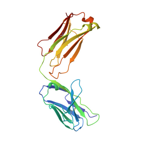
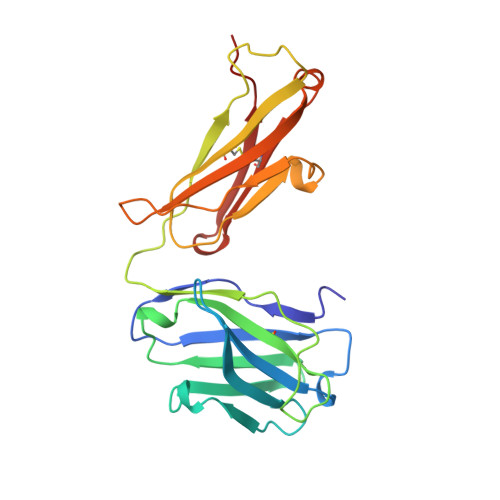
Structural studies of human autoantibodies. Crystal structure of a thyroid peroxidase autoantibody Fab.

Chacko, S.Padlan, E.A.Portolano, S.McLachlan, S.M.Rapoport, B.

(1996) J Biological Chem 271: 12191-12198

  • DOI: https://doi.org/10.1074/jbc.271.21.12191
  • Primary Citation Related Structures: 
    1VGE

  • PubMed Abstract: 

    The three-dimensional structure of the Fab of TR1.9, a high-affinity IgG1, kappa human autoantibody to thyroid peroxidase, was determined crystallographically to a resolution of 2.0 A. The combining site was found to be relatively flat, like other antibodies to large proteins. Sequence differences from the most closely related germline genes mainly occur at positions occupied by residues with outward-pointing side chains. An increased deformability of the second and third complementarity-determining regions of the heavy chain may result from the replacement of two germline asparagines and the presence of several glycines, and may allow "induced fit" in the binding to antigen. Four exposed charged residues, resulting from the use of a particular D (diversity) and J (joining) segments in the assembly of the heavy chain, may contribute to the high affinity of antigen binding. The crystal structure of TR1.9 Fab is the first for a human IgG high-affinity autoantibody.


  • Organizational Affiliation
    • Laboratory of Molecular Biology, NIDDK, National Institutes of Health, Bethesda, Maryland 20895-0560, USA.

Macromolecule Content 

  • Total Structure Weight: 47.18 kDa 
  • Atom Count: 3,534 
  • Modeled Residue Count: 439 
  • Deposited Residue Count: 439 
  • Unique protein chains: 2

Macromolecules

Find similar proteins by:|  3D Structure
Entity ID: 1
MoleculeChains  Sequence LengthOrganismDetailsImage
TR1.9 FABA [auth L]214Homo sapiensMutation(s): 0 
Gene Names: CDNA DERIVED FROM
Find similar proteins by:|  3D Structure
Entity ID: 2
MoleculeChains  Sequence LengthOrganismDetailsImage
TR1.9 FABB [auth H]225Homo sapiensMutation(s): 0 
Gene Names: CDNA DERIVED FROM
UniProt & NIH Common Fund Data Resources
Find proteins for P01857 (Homo sapiens)
Explore P01857 
Go to UniProtKB:  P01857
PHAROS:  P01857
GTEx:  ENSG00000211896 
Entity Groups
Sequence Clusters30% Identity50% Identity70% Identity90% Identity95% Identity100% Identity
UniProt GroupP01857
Sequence Annotations
Expand
Reference Sequence

Experimental Data & Validation

Experimental Data

  • Method: X-RAY DIFFRACTION
  • Resolution: 2.00 Å
  • R-Value Work:  0.180 (Depositor), 0.169 (DCC) 
  • R-Value Observed: 0.180 (Depositor) 
Space Group: P 1 21 1
Unit Cell:
Length ( Å )Angle ( ˚ )
a = 50.926α = 90
b = 62.776β = 107.19
c = 84.79γ = 90
Software Package:
Software NamePurpose
XDSdata scaling
X-PLORrefinement
XDSdata reduction

Structure Validation

View Full Validation Report


Entry History 

Deposition Data

Revision History  (Full details and data files)

  • Version 1.0: 1996-06-10
    Type: Initial release
  • Version 1.1: 2008-03-24
    Changes: Version format compliance
  • Version 1.2: 2011-07-13
    Changes: Version format compliance
  • Version 1.3: 2024-10-23
    Changes: Data collection, Database references, Structure summary